Category Radiation Safety

Is Your Pet Safe During an X-ray?
Illustration of an owner waiting calmly with their pet outside the imaging room

The six questions owners ask while waiting outside the imaging room: is there radiation, can I hug my pet afterwards, how much is one picture, why wait outside, what about pregnancy and children, and do repeat X-rays add up? All answered on one page clinics can share with owners.
Filtration: Removing the Rays That Only Harm and Never Image

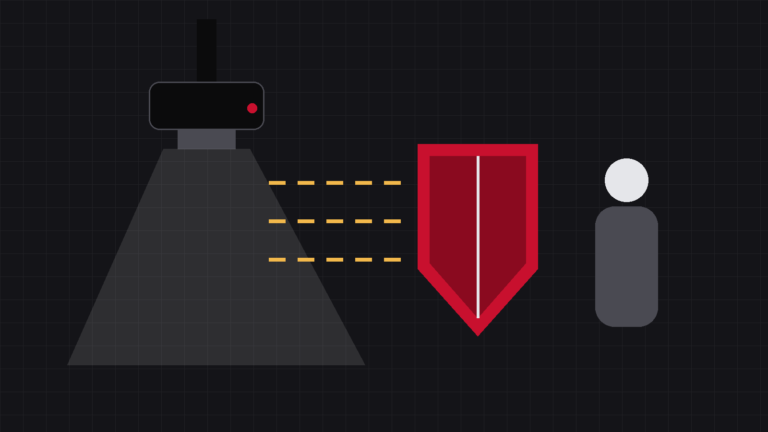
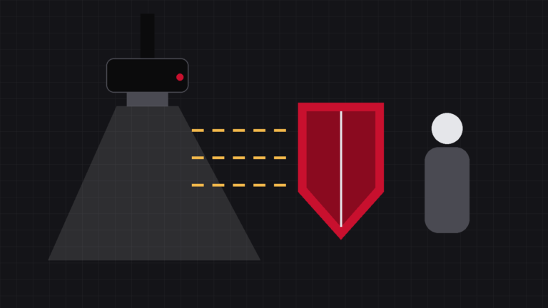
Illustration of an aluminium filter stopping low-energy rays while higher-energy rays pass through

An X-ray tube does not emit one tidy beam; it emits a mixture of energies. The soft part of that mixture cannot get through the body at all — it is absorbed in the skin, the detector never sees it, and the animal receives every bit of it. The remedy is almost primitive: a few millimetres of aluminium across the tube port. Here is where filtration does its work, how inherent and added filtration differ, and three things it means for your clinic — do not remove it, check the half-value layer line, and understand the copper filtration already used in human DR.

Does a Shorter Exposure Mean Less Radiation?

Illustration of a stopwatch next to an X-ray tube

“This machine has a short exposure time, so the radiation is lower” — half true, half not. Total dose follows mAs: 100 mA for 0.1 s and 200 mA for 0.05 s are both 10 mAs, so shortening the time while raising the current saves nothing at all. Where a short exposure genuinely saves dose is elsewhere: the animal has no time to move, the first attempt succeeds, and what you save is the dose of the repeat. Also here: why short exposures depend on generator power, and why “higher kV means more radiation” is not accurate either.

Why DR Uses Less Dose Than Film Did

Illustration of a sheet of film beside a digital flat panel detector

Everyone says DR uses less radiation than film — less where? The position first: this is a difference between generations, not between brands; clinics still on film or CR are using what was the best technology of its day. The saving sits in three places: the detector catches more (higher DQE), latitude rescues near misses (a slightly off technique no longer means a repeat), and the instant image ends the habit of buying insurance with dose. But the dividend does not collect itself — technique chart, exposure index, collimation, first-time-right.

How Much Radiation Does One Radiograph Give an Animal?

Illustration of a dog on an examination table with a large question mark beside it

The question owners ask most, and the hardest to answer honestly. Veterinary medicine has no authoritative dose reference table of the kind human radiology publishes, and patients run from Chihuahua to Great Dane — so if anyone quotes a per-view dose to two decimal places, ask for the source. What is clear are the four factors that decide it: thickness, region, field size and retakes, all of them in your hands. Here is how they rank.

How Thick Does the X-ray Room Wall Need to Be?

Illustration of an X-ray room wall in cross-section with the idea of lead equivalence

“Are my walls thick enough, or do I line the room with lead?” Start with one term: lead equivalence — how much a barrier stops, expressed as a thickness of pure lead. It describes an effect, not a material, so a solid masonry wall of adequate thickness is very often an adequate barrier on its own and most rooms need no lead lining. The actual number comes from your local requirement plus a shielding calculation. Also: the four points where rooms really leak, and the four steps you must not reorder.

Can a Pregnant Technician Be in the X-Ray Room?

Illustration of a technician standing outside the X-ray room door

The answer first: pregnancy does not mean leaving radiography, and it does not mean resigning. International and national rules agree — once the pregnancy is declared, the employer adjusts the working conditions so the dose to the fetus for the rest of the pregnancy stays comparable to that of a member of the public. In a veterinary clinic that comes down to three things: no restraint, out of the room at exposure, role moved up front. Plus the one thing the practice owner should do about it.

Gy, Sv, mSv: What Actually Separates Them

Illustration of three radiation dose unit symbols side by side

A survey report shifts between μGy, mSv and μSv/h and looks like three separate systems. It is not: there are two units and one conversion. The gray is physics and is measured; the sievert is about people and is calculated — and since the radiation weighting factor for X-rays is 1, the two figures line up. Add the steps of a thousand, two everyday reference points, and the rule that a slash means a dose rate, and the table becomes readable.