
Filtration: Removing the Rays That Only Harm and Never Image
An X-ray tube does not emit one tidy beam; it emits a mixture of energies. The soft part of that mixture cannot get through the body at all — it is absorbed in the skin, the detector never sees it, and the animal receives every bit of it. The remedy is almost primitive: a few millimetres of aluminium across the tube port. Here is where filtration does its work, how inherent and added filtration differ, and three things it means for your clinic — do not remove it, check the half-value layer line, and understand the copper filtration already used in human DR.
 Veterinary imaging · Core software built in-house
Filtration: Removing the Rays
That Only Harm and Never Image
What leaves an X-ray tube is not one tidy beam. It is a mixture of energies, and part of that mixture is useless from birth: too weak to get through the body, absorbed in the skin and the tissue just under it. The detector never sees it. The animal receives all of it.
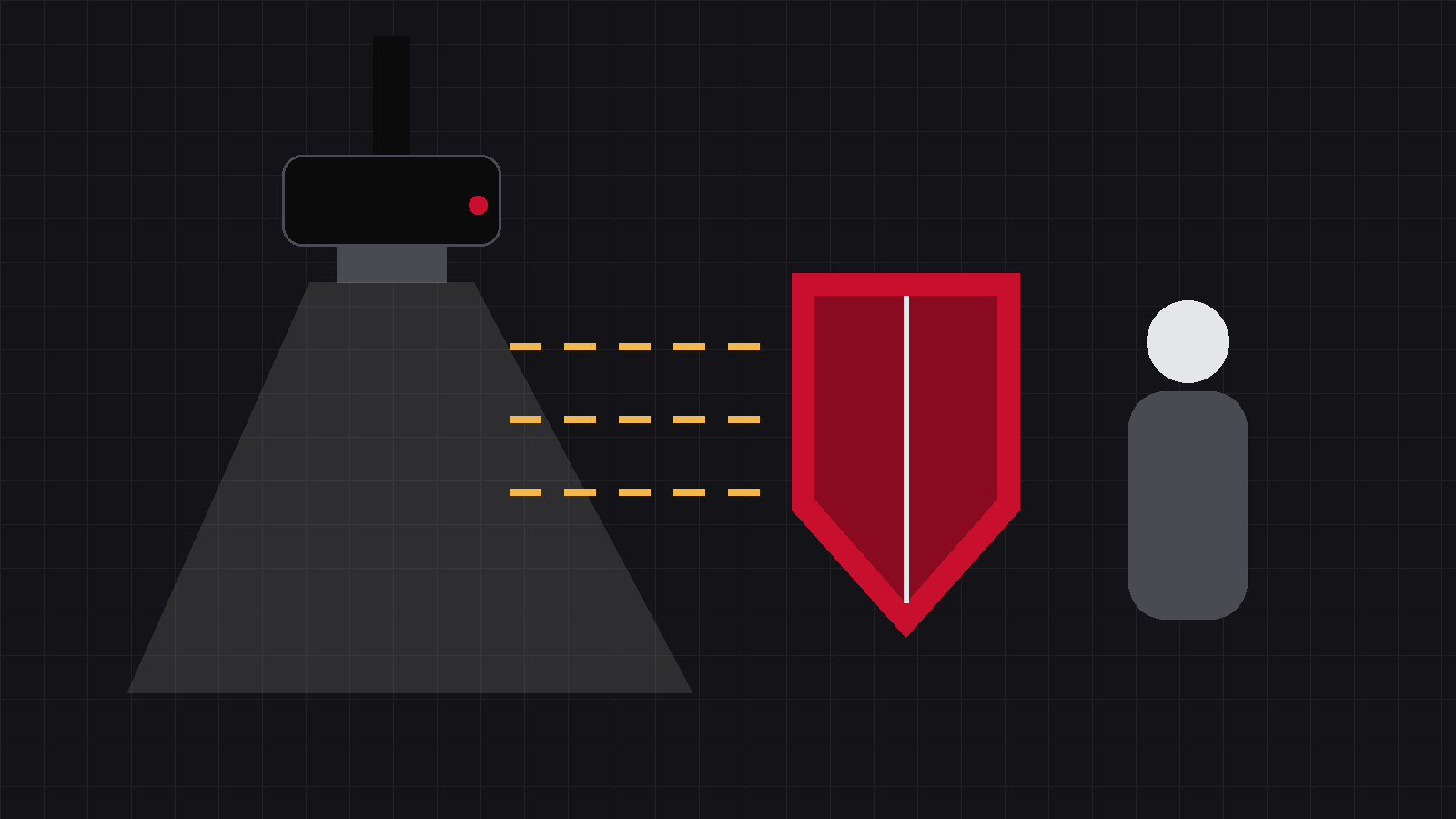
There is one dose-reduction measure your clinic never operates and never sees. It was fitted on the day the machine left the factory, a few centimetres behind the tube port, and it quietly takes a slice off the dose of every image you make. It is called filtration. This issue explains what it does, and why it is worth knowing that it is there.
01A tube emits a mixture, not a beam
We say “the beam” as though it were one uniform thing. What an X-ray tube actually produces is a mixture of photon energies — some high, some low, and everything in between. (Our S2 series went through the tube spectrum in detail.)
Part of that mixture is useless from the start: the energy is so low that it cannot get through the body at all. It is absorbed in the skin and the tissue immediately beneath it. The trade calls these soft rays.
Soft rays are pure harm
They never reach the detector, so they contribute nothing whatsoever to the image. But the place where they are absorbed is the animal’s skin. Zero contribution to the picture, the entire dose on the patient.
02The fix is almost primitive
Put a few millimetres of aluminium across the tube port. The soft rays stay inside the machine; the harder ones go out and do the work. That is filtration.
Why does one sheet of aluminium stop the soft rays and not the hard ones? Because the lower the energy, the more readily a photon is absorbed. To a low-energy photon that aluminium is a wall; to a high-energy one it is a veil. The sheet therefore acts as a sieve, keeping inside the machine the part of the output that was destined to be wasted on skin.
03Filtration comes in two layers
A machine’s filtration is not a single sheet of metal. It is the sum of two things:
Inherent filtration is what the machine has anyway: the glass envelope of the tube, the insulating oil around it, the exit window. None of it was put there to filter the beam, but all of it absorbs some of the soft component, and none of it can be removed.
Added filtration is deliberate: aluminium fitted at or near the collimator. The two together are the total filtration, and the common requirement for diagnostic X-ray units is at least 2.5 mm aluminium equivalent — not an option, a baseline for this class of equipment. Local rules in force may go further in their detail, and local rules are what you follow.
04Why the trade is such a good one
Add filtration, and the entrance dose at the animal’s skin drops appreciably, while the image-forming radiation reaching the detector is barely touched.
Filtration removes only the pure-harm fraction
It does not degrade the image. It takes away the part that was never going to contribute to the image in the first place. Very few dose measures are this clean.
There is one cost. With the soft component gone, the tube must put out a little more in total to compensate, which means slightly more heat load on the tube and anode. That cost is paid by the machine, not by the animal.
Put another way: filtration spends equipment life to buy down skin dose. Equipment can be serviced and replaced. Dose already delivered cannot be taken back.
05What this means for your clinic: three things
① Do not remove it
Added filters do get left out after a service. The dangerous part is this: with the filter missing, the machine still produces images, and the images still look acceptable — while the animal’s skin dose has gone up a step and nobody can see it. So: after any service, confirm that the filtration assembly is back in place. Written on the service sheet beats remembered in someone’s head.
Nothing warns you
No alarm, no error, no visible change in image quality, no change in your technique settings. The only quantity that changed is the one you cannot see.
One question is enough
If the tube or the collimator has been worked on, get the engineer to state plainly whether the filtration assembly was disturbed and refitted — and put the answer on the paperwork.
② It appears in your inspection report
Annual testing usually includes a measurement called the half-value layer: how much aluminium it takes to cut the beam intensity in half. A harder beam needs more aluminium, so this figure is an indirect check that the filtration is adequate. A pass on that line means the radiation coming out of your machine really is filtered.
③ Knowing this, you can read where human radiography is heading
Human DR has for some years added 0.1–0.2 mm of copper filtration on top of the aluminium, cleaning out the soft component further. It is an established dose-reduction technique. The same route is open to veterinary equipment; we will return to it when there is reliable measured data to show.
One last note: issue 7 made the case for re-measuring a room with a survey meter after any service. A machine whose filtration assembly has been disturbed is exactly the kind that earns that measurement.
Seventeen issues into this series, this is the one protective measure you cannot touch and cannot adjust — it is already inside the machine. Your only job is to make sure it does not quietly disappear during a service.
Soft rays harm without imaging.
Filtration stops them at the port — and all of what it stops was dose for nothing.
Source:That an X-ray tube emits a continuous spectrum, and that its low-energy component contributes skin dose without contributing to the image, is standard imaging physics found in radiographic technique texts. A total filtration of at least 2.5 mm aluminium equivalent is the common requirement for diagnostic X-ray units; the specific regulatory clause is whatever is currently in force in your jurisdiction. Half-value layer measurement is a routine item in annual testing and verifies filtration indirectly. Adding 0.1–0.2 mm of copper on top of aluminium is established practice in human DR (documented in the technical literature of the major manufacturers). No brand is named here, and no dose-reduction performance is promised for any equipment.
IWA Medical · We build DR systems and write our own core software
Questions about veterinary DR imaging? Technical questions go straight to our engineers.