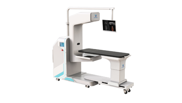
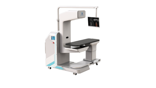
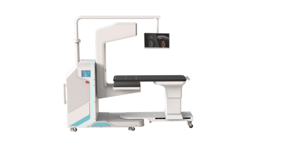
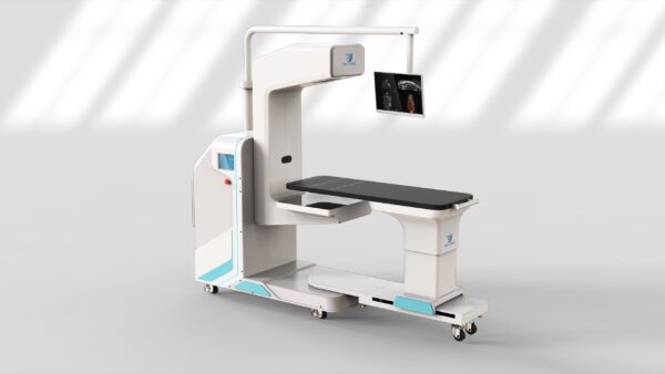
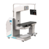
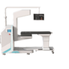
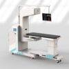
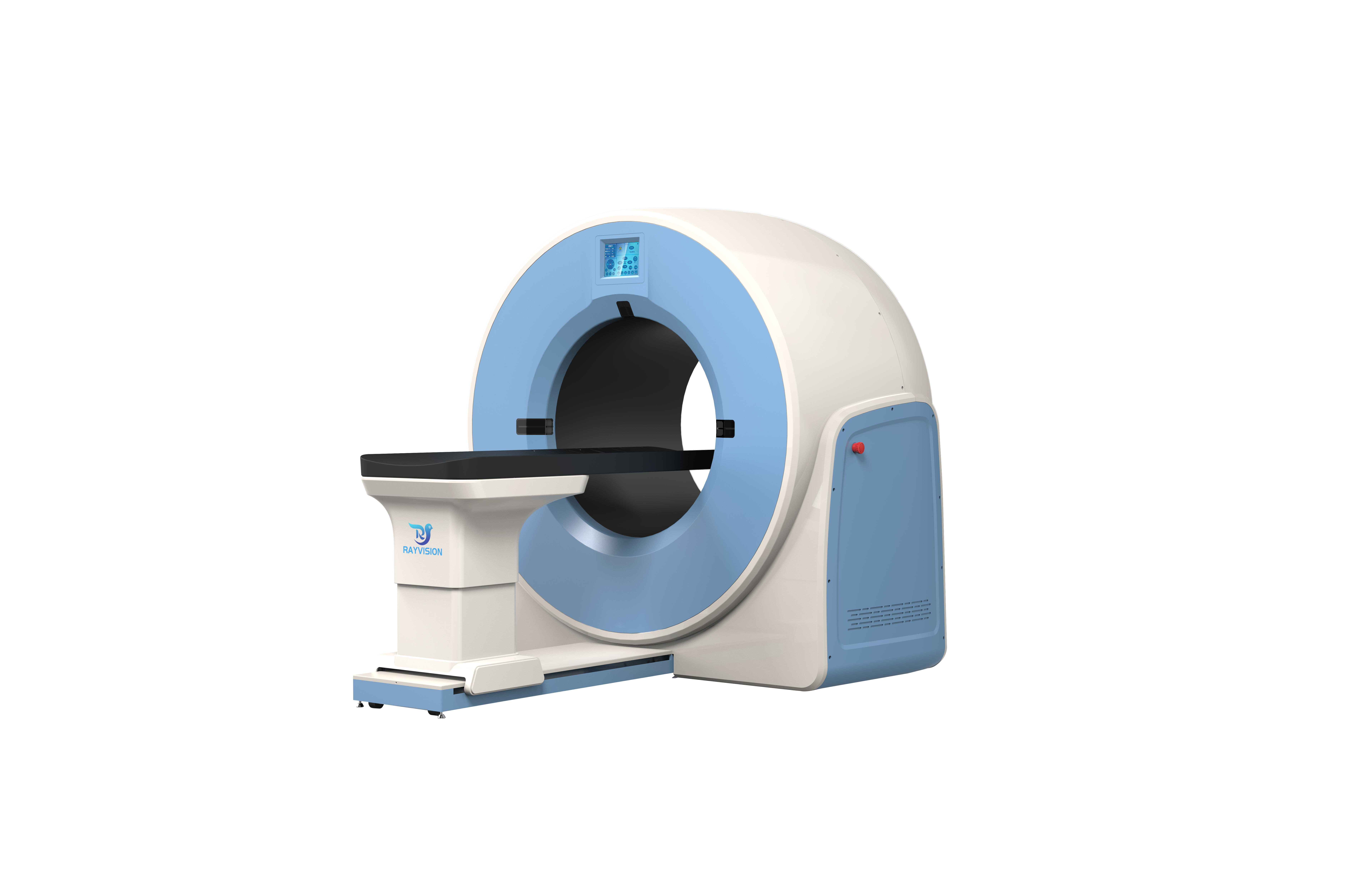
The specialist C-arm CBCT — a 55 × 28 cm field of view, 800 reconstructed slices and dual-mode triple-phase contrast for surgical and interventional caseloads.
Product Overview
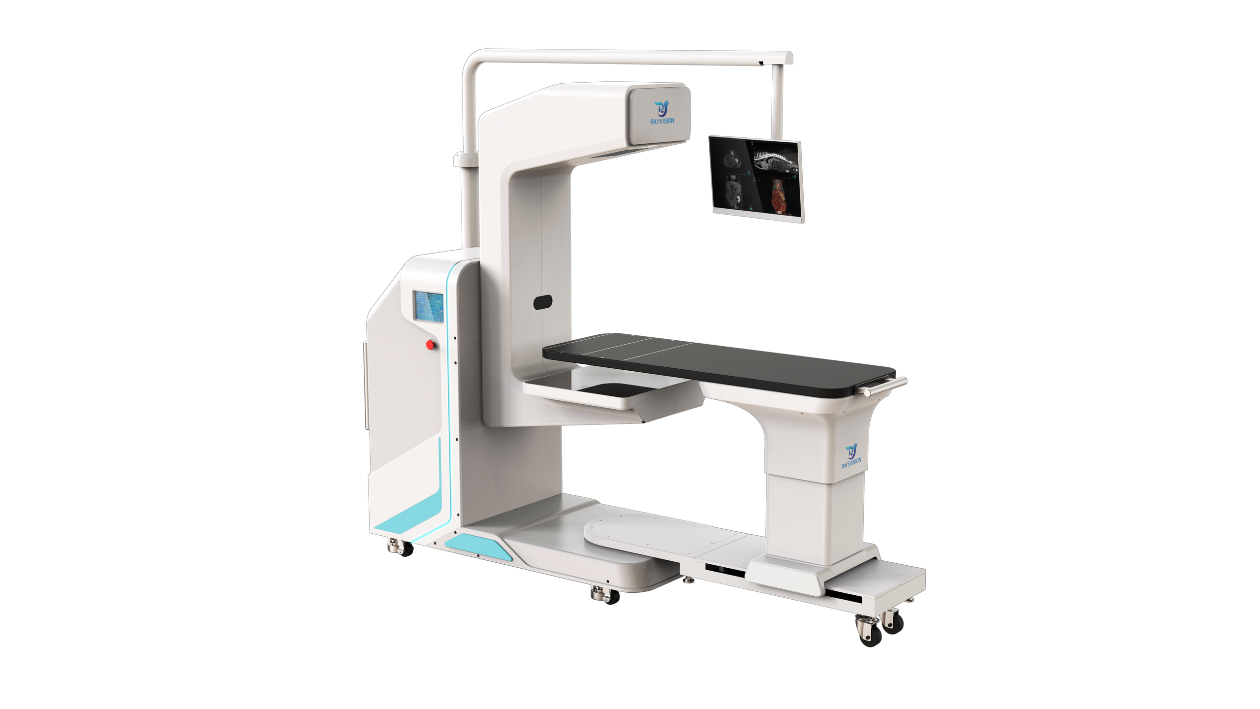
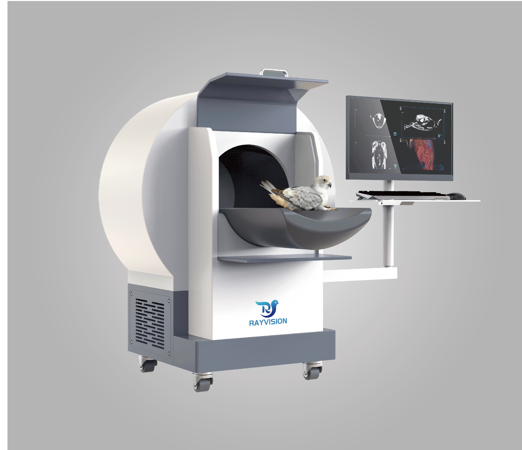
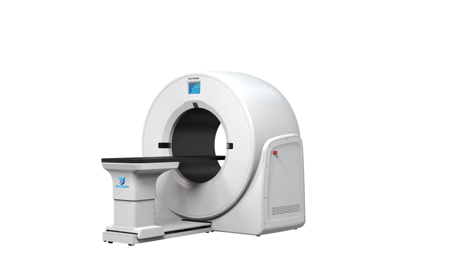
Vetopia 70 (Select Series) is the specialist C-arm veterinary cone-beam CT system from Rayvision Medical. It extends the intraoperative C-arm platform with a 55 × 28 cm CT field of view and 800 reconstructed slices.
Triple-phase contrast capture runs in experience-triggered or time-triggered mode, DSA supports interventional work, and the database-assisted reporting system retrieves similar cases from a built-in library to support diagnosis and report writing.
Key Features
- 55 × 28 cm field of view — 800 reconstructed slices, 0.16 mm minimum slice thickness, 4.5 s scan.
- Dual-mode triple-phase contrast capture — experience-triggered for real-time operator control; time-triggered for preset, synchronised full triple-phase coverage.
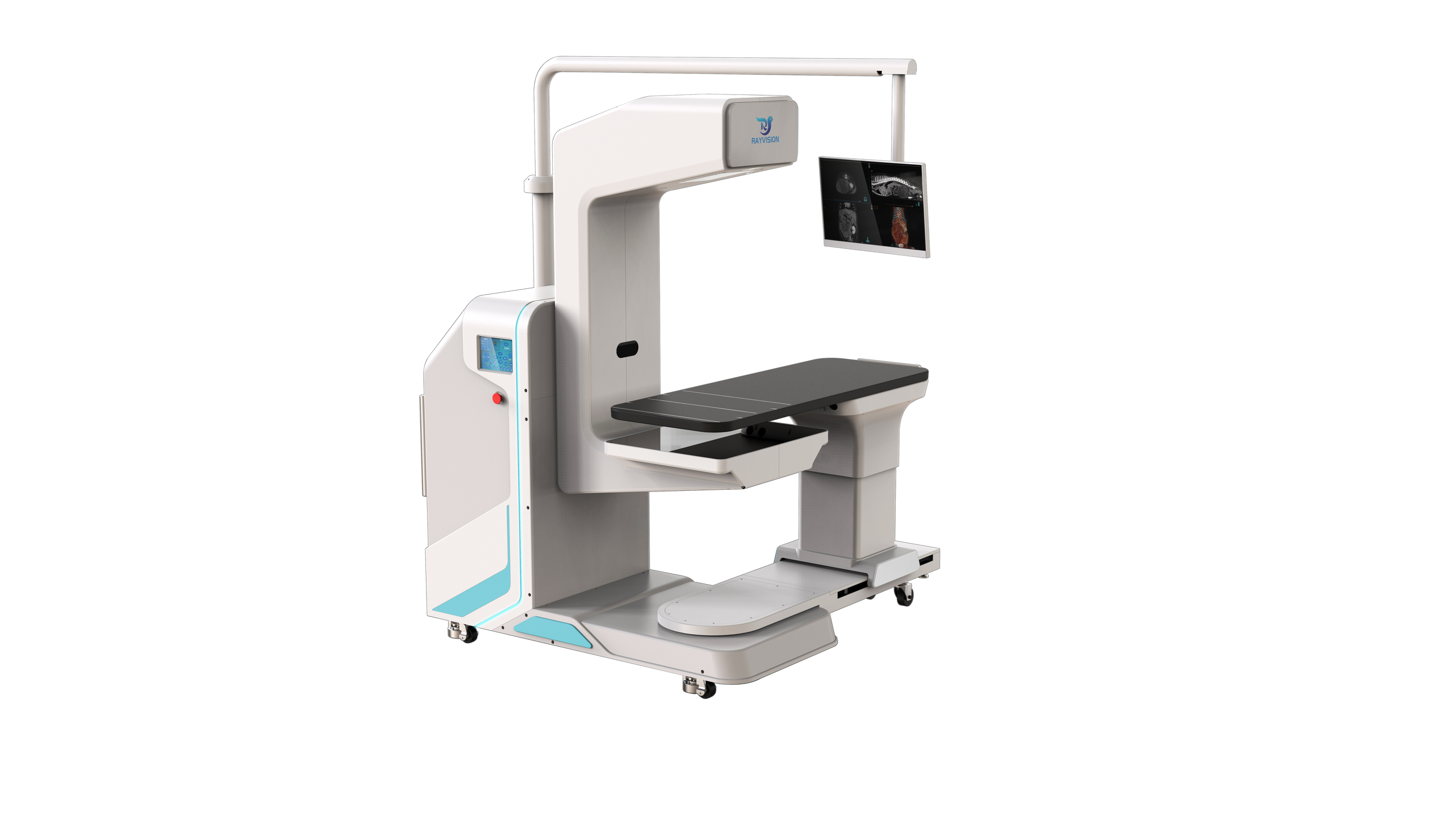
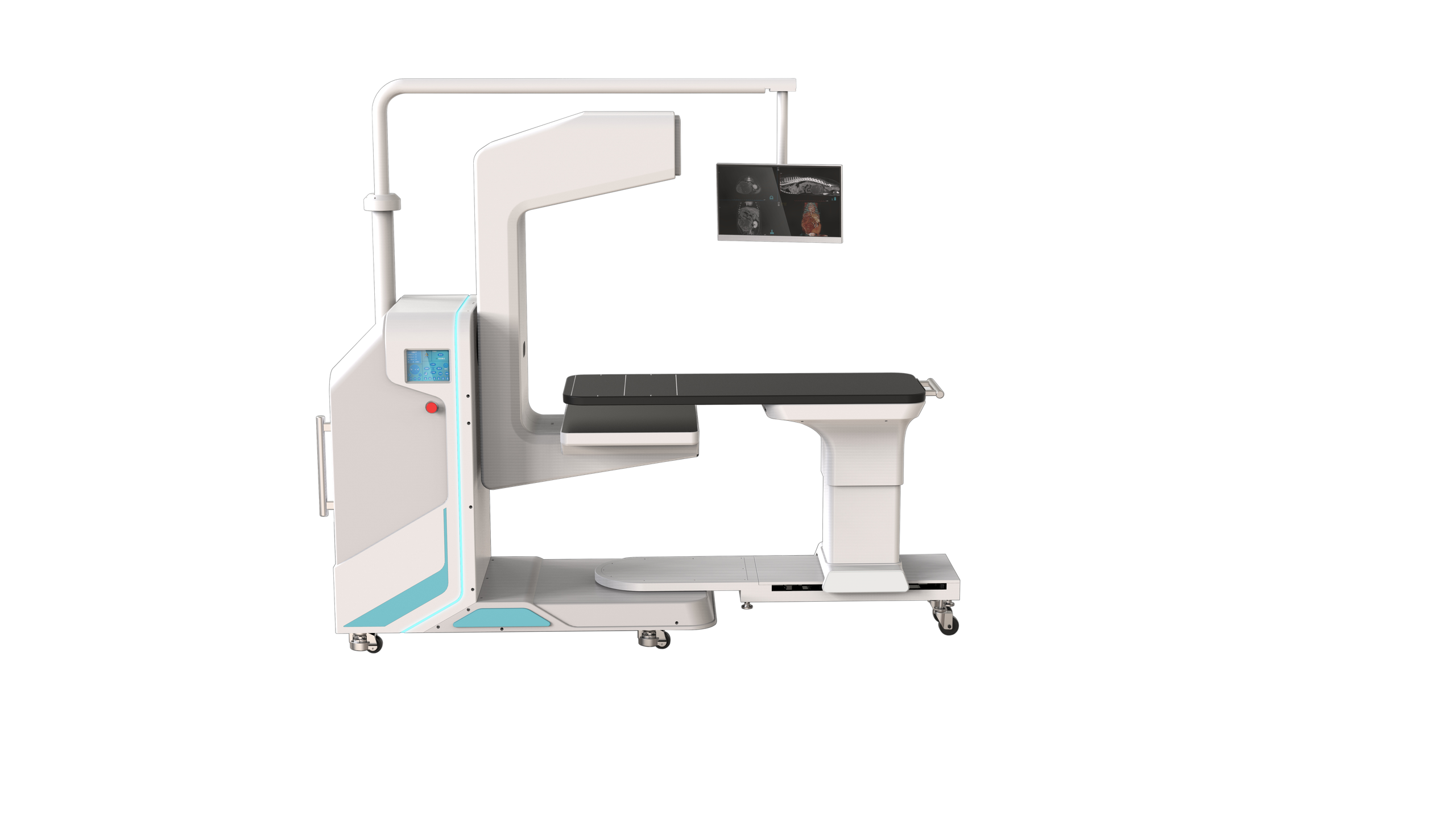
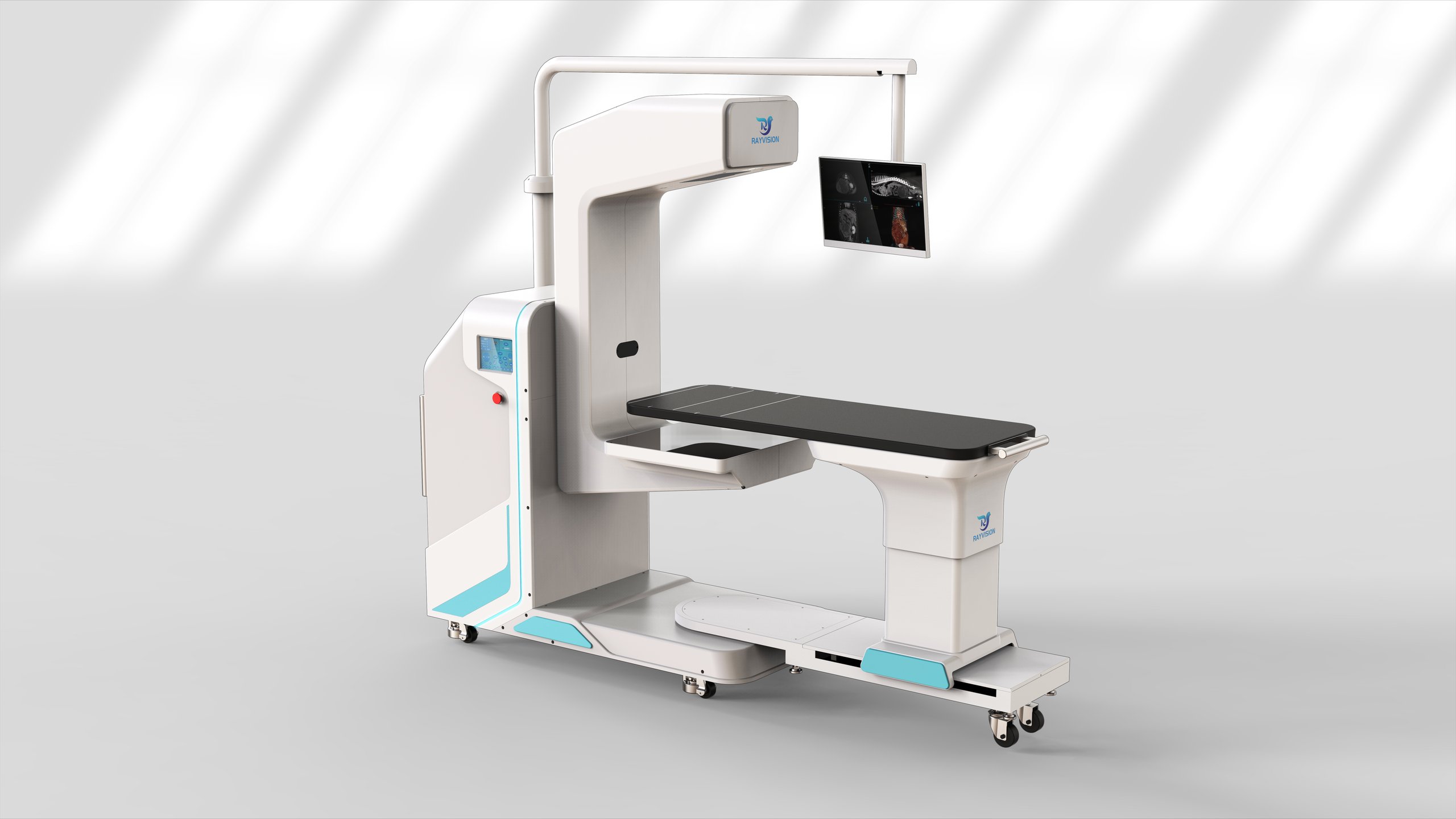
- Flexible C-arm with upgraded rail-mounted table — multi-angle gantry and table rotation; intraoperative monitor at the table side.
- DSA — digital subtraction angiography shows vascular structures to guide interventional procedures.
- Intraoperative dynamic imaging — real-time capture of lesion changes during surgery.
- CT-AIR breath-hold platform — holds the patient at end-inspiration to eliminate motion artefact.
- Smart MAR — suppresses metal artefact around orthopaedic implants for post-operative review.
- Database-assisted reporting — built-in case database for similar-case retrieval and report generation.
Product Gallery
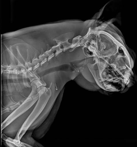
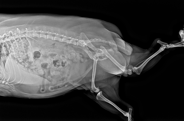
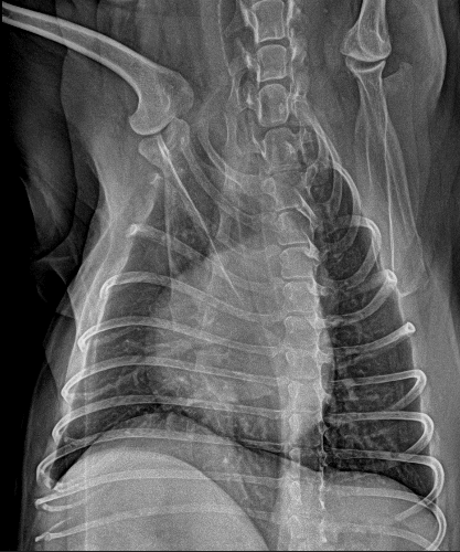
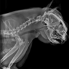
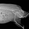
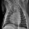
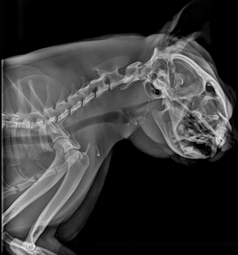
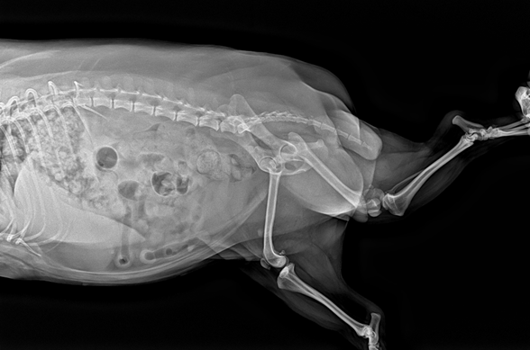
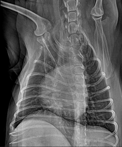
Clinical Imaging Samples
Reference radiographs acquired in DR mode.
Video
Vetopia 70 product introduction
CT case — 21 kg canine patient, nasal tumour
CT case — 6.5 kg feline patient, bronchitis and pneumonia
Technical Specifications — Gantry
| Gantry structure | C-arm (ceiling-rail cantilever) |
| Nominal power | 15 kW |
| Input power | (220 ±22) VAC |
| Tube heat capacity | 150 kJ |
| Maximum tube voltage | 120 kV |
| Tube power | 15 kW |
| Overall dimensions (drawing) | 2446 mm length × 1627 mm height (front view); 678 mm width (side view) |
Technical Specifications — Scan Parameters
| Reconstructed slices | 800 |
| Minimum slice thickness | 0.16 mm |
| Minimum pixel size | 0.14 mm |
| Detector matrix | 3072 × 3072 |
| CT scan time | 4.5 s |
| Maximum FOV (L × W) | 55 × 28 cm (21.65″ × 11.02″) |
Technical Specifications — Imaging Modes
| CT | 0.16 mm slices, CT-AIR breath-hold, dual-mode triple-phase contrast |
| DR | Edge enhancement, multiple processing algorithms, TPLO / VHS / TTA measurement tools |
| Fluoroscopy (dynamic DR) | Real-time dynamic enhancement, up to 1 minute continuous exposure |
| Dental panoramic | Full upper and lower arches, jaw and temporomandibular joints from one scan |
| DSA | Digital subtraction angiography for interventional guidance |
| Post-processing | Smart Imaging Enhancement algorithm suite |
Clinical Applications
| Region | Conditions |
|---|---|
| Head & neck | Cerebral oedema, brain haemorrhage, sinusitis, otitis, mastoid effusion, nasopharyngeal stenosis, tracheal narrowing |
| Dental | Periodontitis, periapical abscess, alveolar bone atrophy, oral cysts, tooth root resorption |
| Thoracic | Pulmonary oedema, pulmonary nodules, interstitial pneumonia, pulmonary inflammation |
| Abdominal | Abdominal wall inflammation, peritoneal effusion, urography, renal imaging, cholelithiasis, pancreatitis, renal sclerosis |
| Vascular | Abdominal triple-phase angiography, peripheral arterial thrombosis, vascular stenosis |
| Spinal | Myelography, disc protrusion, vertebral imaging, spondylosis, thoracic vertebral fracture |
| Oncology | Thyroid, nasal, ovarian, renal and adrenal tumours |
| Other | Arthritis, thoracic duct imaging, peritoneal calcification |
Clinical Case Highlights
- Renal infarction — Plain scan of the right kidney was unremarkable; after contrast, a cortical enhancement defect indicated partial right renal infarction.
- Gallbladder mucocele — Homogeneous hepatic enhancement in the delayed phase; the gallbladder was filled with irregular soft-tissue-attenuating material with uneven wall thickening.
- Post-processing — Cardiac 3D rendering, vascular MIP and curved reconstruction of the thoracic duct.
Site Planning and Installation
Vetopia 70 is a fixed installation in a shielded room. The manufacturer’s layout drawing uses a 3000 × 3000 mm room with an 850 mm door.
Power and Environment
| Supply voltage | Single-phase (220 ±22) VAC |
| Supply internal resistance | ≤ 0.5 Ω |
| Frequency | 50 Hz / 60 Hz |
| Power capacity | ≥ 15 kVA |
| Power cable | 6 mm² |
| Shielded room | 3 mm lead equivalent |
| Operating temperature | 10 °C – 35 °C |
| Relative humidity | 30 % – 75 % RH, non-condensing |
| Atmospheric pressure | 700 – 1060 hPa |
| Wall clearance | ≥ 30 cm around the system |
Shielded Room Preparation
Room requirements from the manufacturer’s CT installation-conditions guide; a site survey confirms the final layout.
| Room area | Minimum 3 × 3 m; 3.5 × 3.5 m recommended |
| Room height | ≥ 2 m |
| Door opening | ≥ 1 m wide for whole-unit entry (≥ 0.85 m with the main-unit covers removed); ≥ 1.8 m high |
| Wall shielding | 240 mm clay brick + 20 mm protective coating, or any construction equal to 3 mm lead; concrete walls ≥ 240 mm |
| Floor / ceiling shielding | Ground floor: ceiling 3 mm Pb. Intermediate floors: floor and ceiling 3 mm Pb. Top floor: floor 3 mm Pb |
| Floor | Level concrete, foundation depth > 120 mm, flatness ≤ 2 mm, load capacity > 500 kg/m²; ground floor preferred |
| Delivery route | Ramps < 5°; use a forklift or lifting equipment for steeper access |
| Observation window | Standard lead glass 400 × 600 × 15 mm in a 405 × 605 mm opening, supported by 40 × 60 mm square tube |
| Cable entry | Ø 50 mm hole near the operator console |
| Electrical | Dedicated circuit breaker; voltage stabiliser if mains deviates more than ±10 %; UPS where outages are frequent |
| Signage | Radiation warning sign and exposure indicator light outside the room door |
Workflow Notes
- Choose experience-triggered capture for real-time control of each phase, or time-triggered capture for a preset, synchronised protocol.
- The database-assisted reporting system retrieves similar cases and fills report templates from structured data, reducing manual entry.
- The C-arm, rail-mounted table and table-side monitor are positioned around the surgical field.
Package Contents
- 1 × C-arm gantry
- 1 × Rail-mounted multi-angle patient table
- 1 × Intraoperative monitor
- 1 × Acquisition and reconstruction workstation
Downloads
Product brochures and technical documentation are available on request — contact IWA.
Why Choose Vetopia 70
- Workflow efficiency — Time-triggered triple-phase protocols and database-assisted reporting cut manual steps from contrast studies and report writing.
- Clinical accuracy — A 55 × 28 cm field of view, DSA and intraoperative dynamic imaging support complex surgical and interventional cases.
- Practice economics — One C-arm system covers intraoperative CT, fluoroscopy, DR, DSA and dental panoramic imaging for specialist caseloads.
Warranty and Support
1-year whole-system warranty. Extended warranty is available for purchase — contact IWA for terms and service coverage in your region.
Installation is carried out against the manufacturer’s installation manual: the main unit and patient table are connected, the console and workstation are cabled, and scanning, reconstruction, fluoroscopy and DR acquisition are verified before handover.
FAQ
Q: How does Vetopia 70 differ from Vetopia 50?
Vetopia 70 has a 55 × 28 cm field of view (Vetopia 50: 28 × 28 cm), 800 reconstructed slices (Vetopia 50: 600), time-triggered as well as experience-triggered triple-phase capture, and database-assisted reporting.
Q: Is it suitable for interventional procedures?
Yes. The C-arm design, DSA and intraoperative dynamic imaging are intended for surgical and interventional guidance.
Q: What room does it need?
A shielded room of 3 mm lead equivalent (manufacturer layout: 3000 × 3000 mm), a single-phase (220 ±22) VAC supply of ≥ 15 kVA on a dedicated breaker, and 30 cm clearance around the system.
Q: What does database-assisted reporting do?
It retrieves similar cases from a built-in database and fills report templates from structured data, reducing manual entry.
Q: What warranty applies?
1-year whole-system warranty, with extended warranty available for purchase.
Reviews
There are no reviews yet.