VHS
Vertebral heart score
Canine 8.5–10.5 · Feline 7.0–8.0
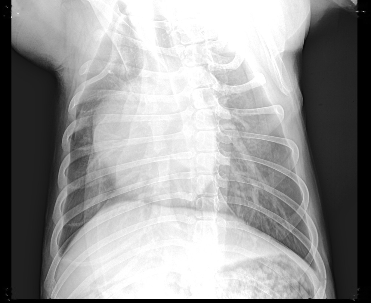
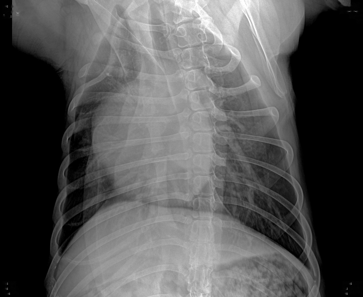
Same panel, same exposure — a different processing algorithm puts the image in a different class. The iPet imaging algorithms are written in-house, specifically for canine and feline radiography: from a 2 kg cat to a 60 kg giant-breed dog, one parameter set delivers the image.
Developed by IWA Medical · veterinary DR system manufacturer
 Before
After
Before
After
Drag the divider. All six cases are real acquisitions — both sides are the same exposure of the same study: the raw capture on the left, the algorithm's output on the right.
Human radiology deals with patients of similar size in standardised positions. Veterinary radiology does not — the same system images a 2 kg cat today and a 60 kg giant breed tomorrow, and on a single canine lateral thorax the penetration across lung field and spine differs by an order of magnitude. Stretch that with one fixed curve and either the lungs disappear or the bone burns out white. Our algorithms were rewritten from that starting point.
The image is decomposed into multiple scales and processed per scale: large scales compress thickness differences so thorax and abdomen read on one screen; small scales pull out trabecular bone and lung texture while noise is suppressed per scale — detail comes up without the noise coming with it.
Every detector model has its own response curve; generic parameters are always off. We calibrate per panel model: bone density and thin soft tissue are each anchored to a target grey level, so changing panels needs no re-tuning. The panel identity travels with the image — copy a case to another workstation and the processing result stays identical.
Grid studies often carry a fine line pattern. The algorithm locates the grid peaks in the frequency spectrum and removes them point by point, second harmonics included. If no grid pattern is found, the image is returned untouched, pixel for pixel — better no processing than invented changes to image data.
On the right is a real radiology step-wedge phantom — a row of aluminium steps of increasing thickness. Drag to change the display window: the narrower the window, the better tissues of similar density separate; open it too wide and everything turns to grey mush. Windowing is the first step of reading a study — and iPet computes the window width and level automatically for every processed image, so the clinician does not have to drag each one by hand.
Algorithms are not "tuned and done". Every change is re-run offline against the same corpus of real raw acquisitions and compared study by study; a version ships only when not a single image got worse — including the hard criterion that an unchanged pipeline must reproduce the previous version pixel for pixel.
Registration, positioning, exposure, processing, measurement, and reporting all happen on the same workstation — each step's output feeds the next. Every vendor claims a workflow; ours is simply made to run smoothly.
Enter patient and owner details; picking a body region expands its views, each card carrying its positioning diagram and exposure preset. Shoot-first, register-later is also supported.
While waiting for exposure, the main display shows the standard positioning diagram — 105 canine and feline views are built in, and the selected view is the one displayed.
kVp / mA / ms / mAs are set automatically by species, body weight, and view. If parameter download fails, exposure is blocked — no image is ever taken on stale parameters.
The raw image appears first for positioning check; enhancement runs on a background thread with visible progress, then window width and level are computed and applied automatically.
16 veterinary measurements with step-by-step guidance, results with reference ranges; anatomically impossible values are flagged invalid and never drawn onto the image.
Templates and standard phrases, an optional AI draft of the findings; the veterinarian verifies line by line before sign-off, and key actions are written to the audit log.
The three screenshots below are taken from a running system. The interface is dark by design: reading rooms are dim, and a bright interface both glares and skews greyscale judgement. Functions sit directly on the page, not buried in nested menus.
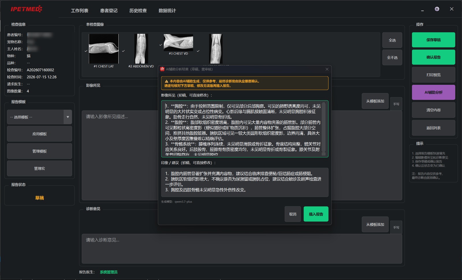
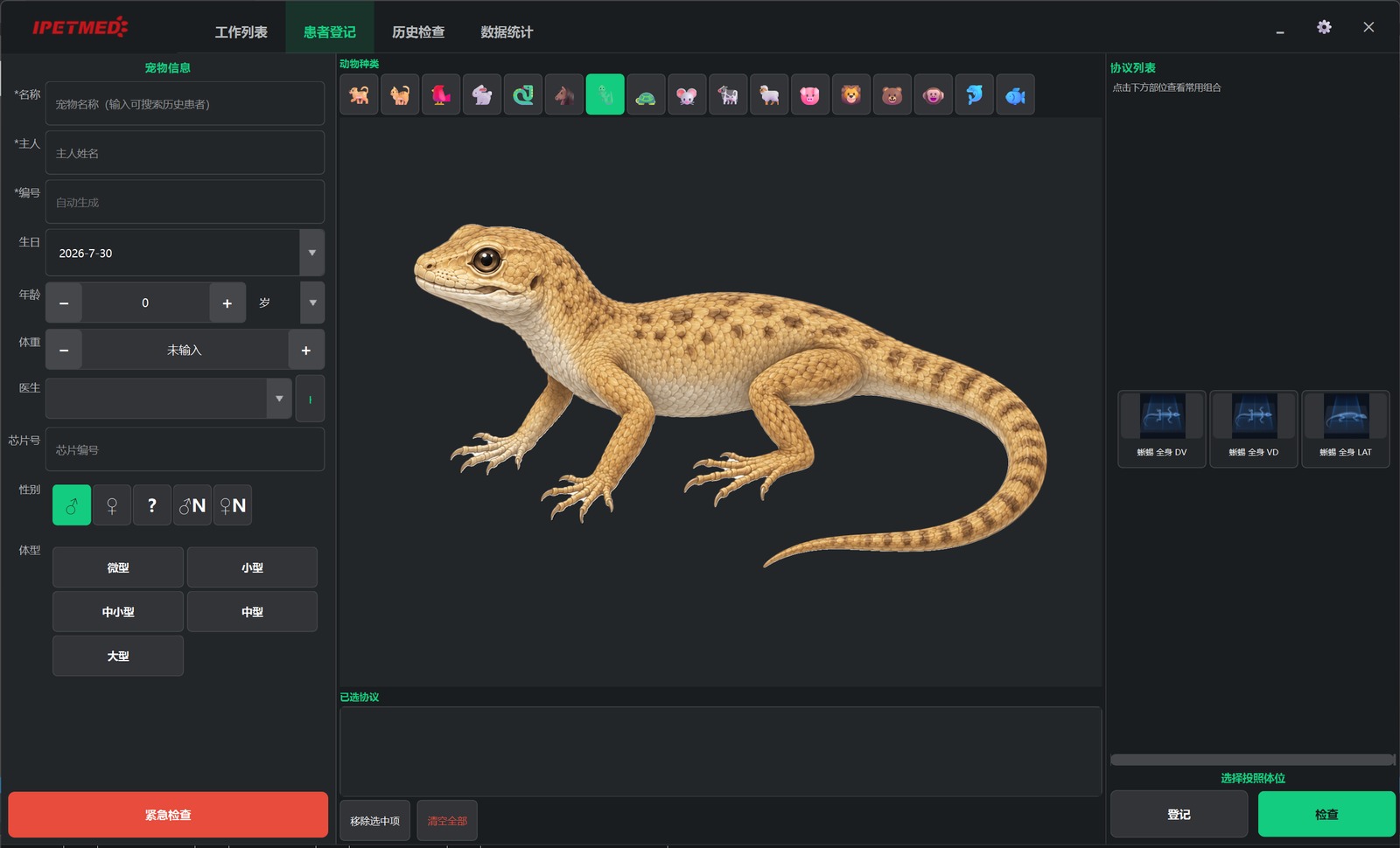
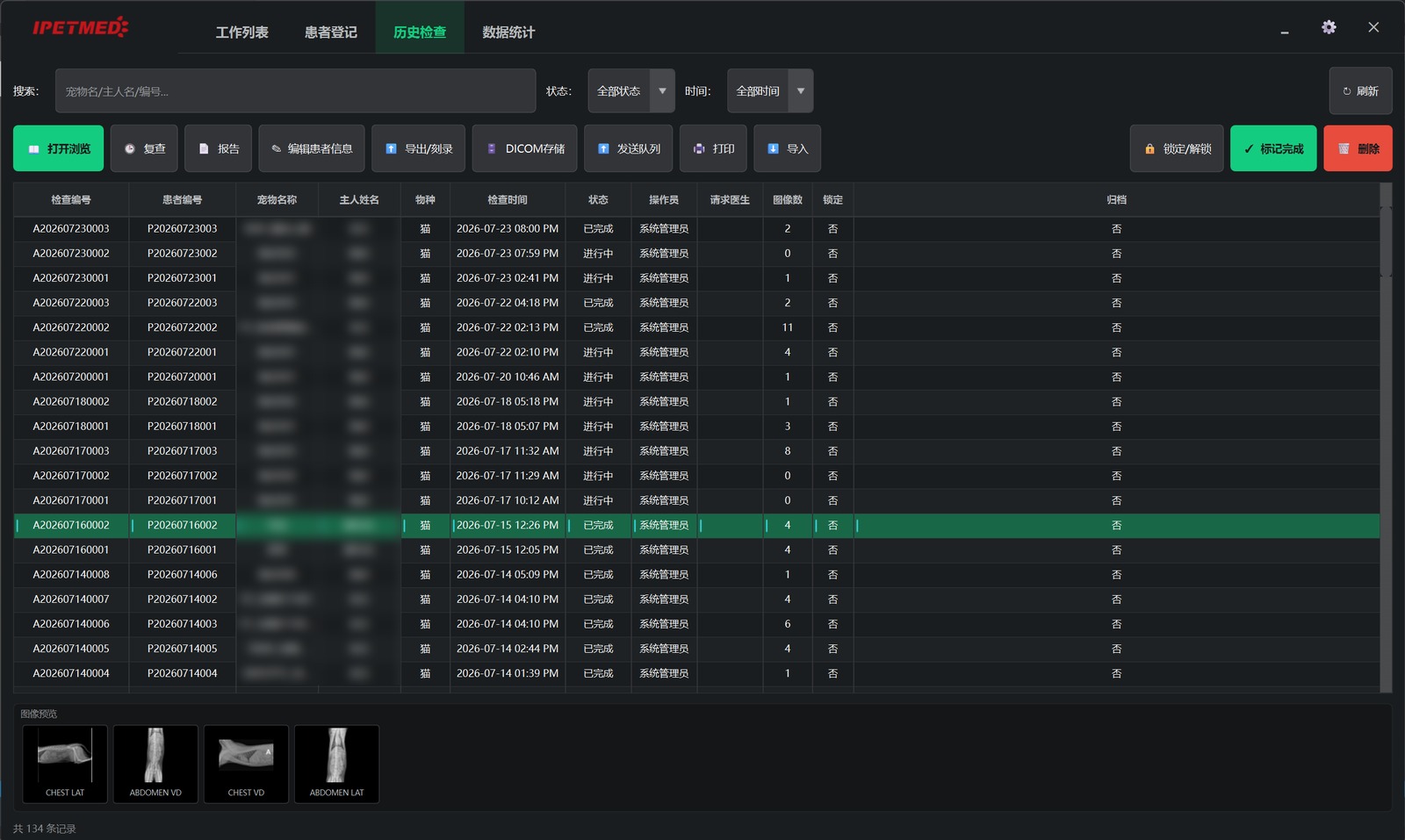
Screenshots are from a development validation system with pet and owner names blurred; the interface is shown in Simplified Chinese here and ships in 8 languages, English included.
An image starts earning its keep after it is taken — and that is where many hospitals get stuck: studies live only on the acquisition PC, clinicians cannot see them in the consult room, referrals travel by USB stick, and owners photograph the screen. We built the whole chain — and all three products are ours, so no third party has to cooperate on the interfaces.
The machine in the imaging room. Every study is generated to the DICOM standard, archived locally, and pushed to the in-hospital server automatically.
Installed on the hospital's own server, receiving studies from every modality in the building. Data never leaves the hospital — any PC in the consult room, ultrasound room, or director's office reads studies in a browser.
For reading across branches and locations. Once studies are in the cloud, practice management, medical records, prescriptions, billing, and imaging run in one connected system.
The three tiers can be bought separately or singly. Any DICOM-conformant device can push studies — enter server address, port, and AE title. Nothing is locked to our own systems.
Cardiac scoring, hip evaluation, osteotomy planning — the metrics canine and feline clinicians actually use, and which human-medicine software simply does not have. All built in; no plug-ins to buy.
For measurements needing seven or eight landmarks — VHS, PennHIP, TPLO — the wizard shows which point to place next and which anatomical landmark it is. No memorising sequences.
Each value comes with the normal range for the species — VHS canine 8.5–10.5, feline 7.0–8.0 — no digging through the literature to compare.
If mis-placed landmarks produce an anatomically impossible result, the software flags it invalid and explains the correct placement — a misleading number is never drawn onto the image.
For true millimetre output, run a one-time distance calibration: draw a line of known length on the image and enter the real dimension. Ratio metrics such as VHS need no pixel spacing — a reference vertebra is calibrated per the standard method.
Veterinarians do not lack reading skill; they lack the time to turn what they see into a structured report. That is where the AI earns its place: it writes a first draft in the standard structure, and editing a draft is far faster than starting from a blank page.
The current study, together with species, region, and clinical notes, goes to the iPet AI reading model, which returns a draft in a two-part "Findings / Impression" structure. The model is explicitly constrained: state uncertainty as uncertainty, never invent measurement values, never prescribe treatment.
The cloud platform analyses uploaded studies and cross-checks the AI's output against six rule classes — species mismatch, sex conflict, age conflict, disease blacklist, wrong body region, internal consistency. Flagged passages are marked with red or amber warning bars — telling the clinician exactly where the AI may be wrong.
Clinicians can rate every AI reading, and the system aggregates the ratings into accuracy statistics. The longer it runs, the clearer it becomes where the AI can be trusted — and where it cannot.
—
—
This content is AI-assisted and for reference only; the final diagnosis must be confirmed by a licensed veterinarian.
↑ The section structure and disclaimer are the software's actual output format; the text is illustrative.
From 1.0 to 2.0.2, the capabilities below shipped in the last two months and are running in hospitals now. All updates arrive through in-app automatic upgrade, with patient data and settings preserved as they are.
Every acquisition passes an AI quality check first: exposure adequate, positioning correct, motion artefact present or not — failures come with the reason. Processing parameters fine-tune themselves against per-region baselines: the more you shoot, the closer they get.
Bone and soft tissue emphasised in separate layers, with a one-click pseudo-colour mode for soft-tissue density differences — layers the eye cannot separate in thorax or abdomen, colour separates for you.
Severely underexposed studies are auto-windowed for rescue so a retake is often unnecessary; the collimator's lead frame is detected automatically so it no longer pollutes the histogram and bone is not dragged to white.
Pull filtered worklists from the practice system, query and retrieve priors, receive outside studies, report MPPS and storage commitment; RDSR dose reports sent on demand.
A patient's prior studies open side by side automatically, with zoom and windowing linked. Fracture healing and post-operative rechecks no longer mean flipping between two windows.
USB or disc exports carry a no-install viewer, or generate a QR-code link — owners and referral hospitals read studies without installing anything.
Daily automatic backup of database, settings, and image increments with one-click restore; password rules and lockout on repeated failures; DICOM exports can strip owner and patient identity for external consults or publication.
Every exposure's radiation dose is logged automatically, exportable as a safety report at any time; on a device error, the software presents the handling steps and can report to an IWA engineer in one click.
Film printing over the DICOM standard; long-bone and spine series stitch into full-length images; the interface ships in 8 languages including English, with remote assistance built in.
The version number is at the bottom right of the software. Hospitals on 1.0.x are prompted to upgrade to 2.0 automatically — the whole path needs no reinstall and no panel re-calibration.
IWA Medical is a veterinary DR manufacturer, not a software reseller. Flat panels and generators are integrated by us directly at SDK and protocol level — no third-party finger-pointing at installation.
| Flat-panel detector | In-house, high resolution, high speed, ruggedised |
|---|---|
| HV generator | Co-developed with a leading generator manufacturer; matched to the software |
| Acquisition modes | Manual exposure · AED automatic exposure detection · dynamic fluoroscopy (phase 1) · multi-image stitching |
| Imaging standards | DICOM read/write · C-STORE · C-FIND / C-MOVE query-retrieve · SCP receive · Worklist · MPPS · storage commitment · RDSR dose report |
| Output | Local export · ISO image / disc burning · DICOM film print · PNG / JPG (with corner overlays) · owner delivery package (no-install viewer / QR link) |
| Environment | Windows desktop; memory- and thread-optimised for entry-level all-in-ones (N100 class); interface in 8 languages |
| Security & compliance | Role-based users · password rules and login lockout · audit log of key actions · daily automatic backup with one-click restore · de-identified export · radiation dose ledger |
The iPet workstation software is distributed through authorised dealers. Dealers download the latest installer after signing in on this site; hospitals should contact us or their supplier.
Sign in with your dealer account and pick up the latest installer on the downloads page. File checksums are published alongside, so the package can be verified before installation.
Go to downloadsThe software ships with the system, installation and commissioning included. To upgrade an existing installation, or to see it running first, get in touch directly.
Mark@iwamed.comPhone +86 134 2443 8112
Installed workstations never need to visit the site. The software checks for new versions at startup; one click upgrades, with patient data, study records, and settings all preserved.
The fastest evaluation is with real cases: shoot a few images on your existing panel, or import prior DICOM studies, and judge the enhancement and measurement workflow for yourself. Full systems, software-only licensing, and upgrades of existing equipment are all on the table.
Phone +86 134 2443 8112 Email Mark@iwamed.com Web www.iwamed.com
This software is an image acquisition, processing, and management tool; measurement values and reference ranges are provided for clinical reference. All diagnostic conclusions are made by a licensed veterinarian.