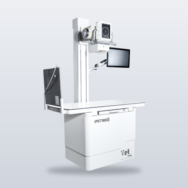
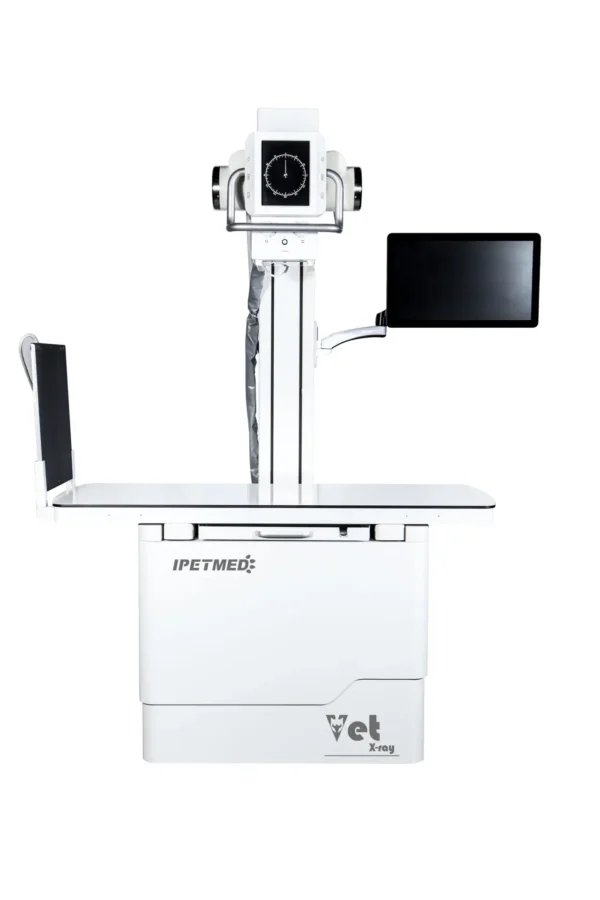
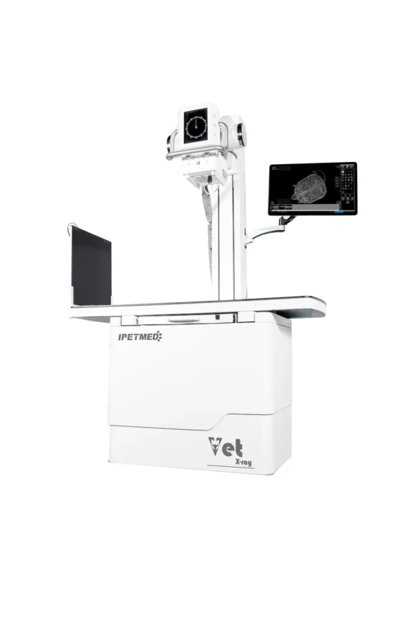
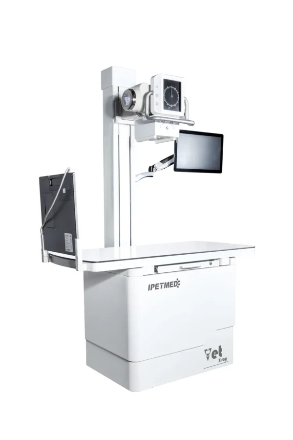
A DR platform built not to be outgrown — iPet-400Pro, the upgradeable static flagship of the iPet AI-DR line.
Product Overview
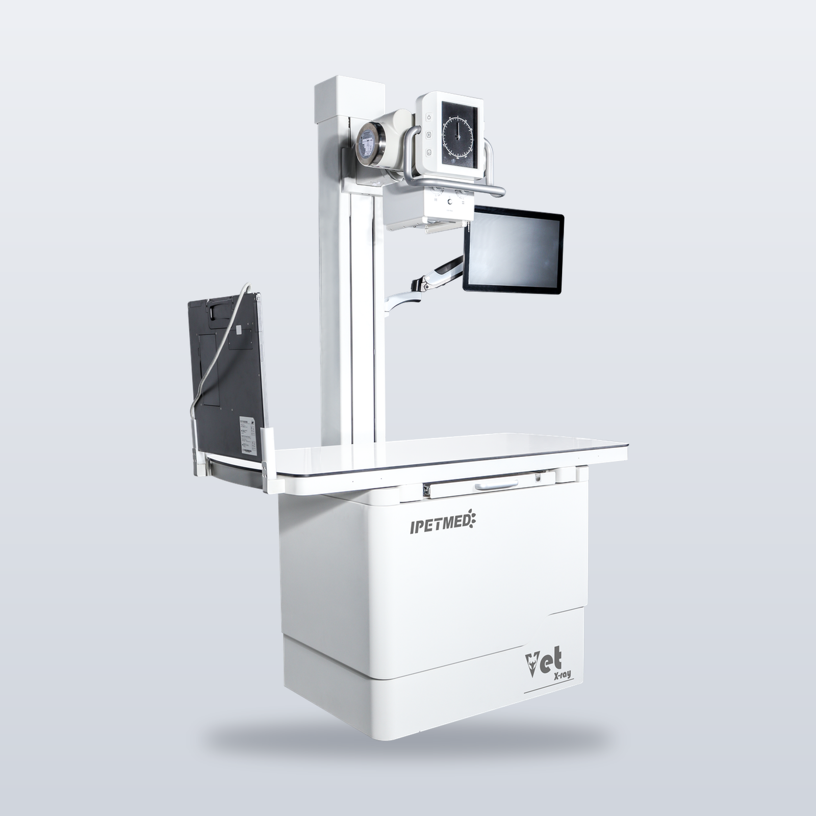
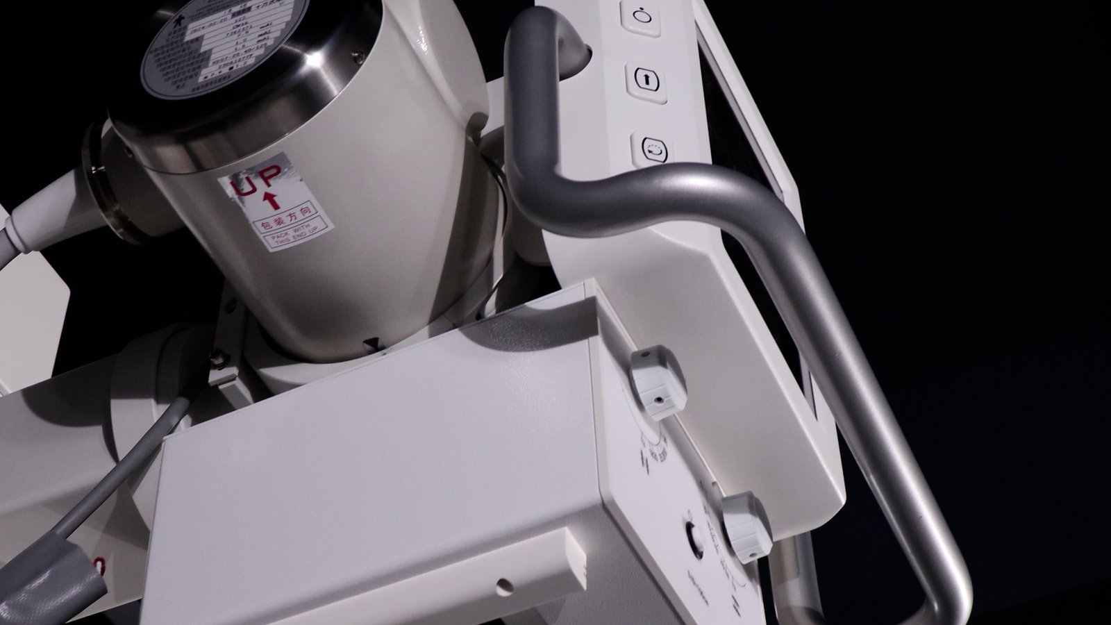
Most DR systems are fixed in configuration the day they leave the factory. iPet-400Pro is not. It is built on the iPet Pro platform: an elevating, rotating gantry lets the tube travel vertically and rotate 90° for horizontal-beam projection, so recumbent, standing, and natural-posture views can all be completed by a single operator. The Canon E7240X tube sustains high-throughput exposure days; the iLume 4343 flat panel is tuned at SDK level with the in-house acquisition software (an original iRay Venu 1717XV panel with verifiable serial number is available as an upgrade option).
More importantly, the platform leaves the door open: reserved upgrade paths for the detector, tube, and high-voltage generator mean today’s static flagship can be upgraded to a dynamic DR configuration later — protecting the practice’s initial investment. The Pro software package adds AI report drafts and AI lesion overlay display.
Key Features
- Elevating, rotating gantry — 90° horizontal-beam projection for standing and natural-posture views, positioned by a single operator.
- Upgradeable platform — reserved upgrade paths for panel, tube, and generator; static configuration can be upgraded to dynamic without replacing the system.
- Canon E7240X tube — 0.6 / 1.2 mm dual focus for fine bone detail and high-throughput work; iLume 4343 flat panel as standard (iRay Venu 1717XV optional, serial-verifiable).
- Pro software package — AI report drafts with veterinarian sign-off, plus AI lesion overlay display.
- DICOM worklist — order straight to DR — exam orders created in practice-management software arrive at the DR workstation automatically; no double data entry from reception to imaging.
Product Gallery
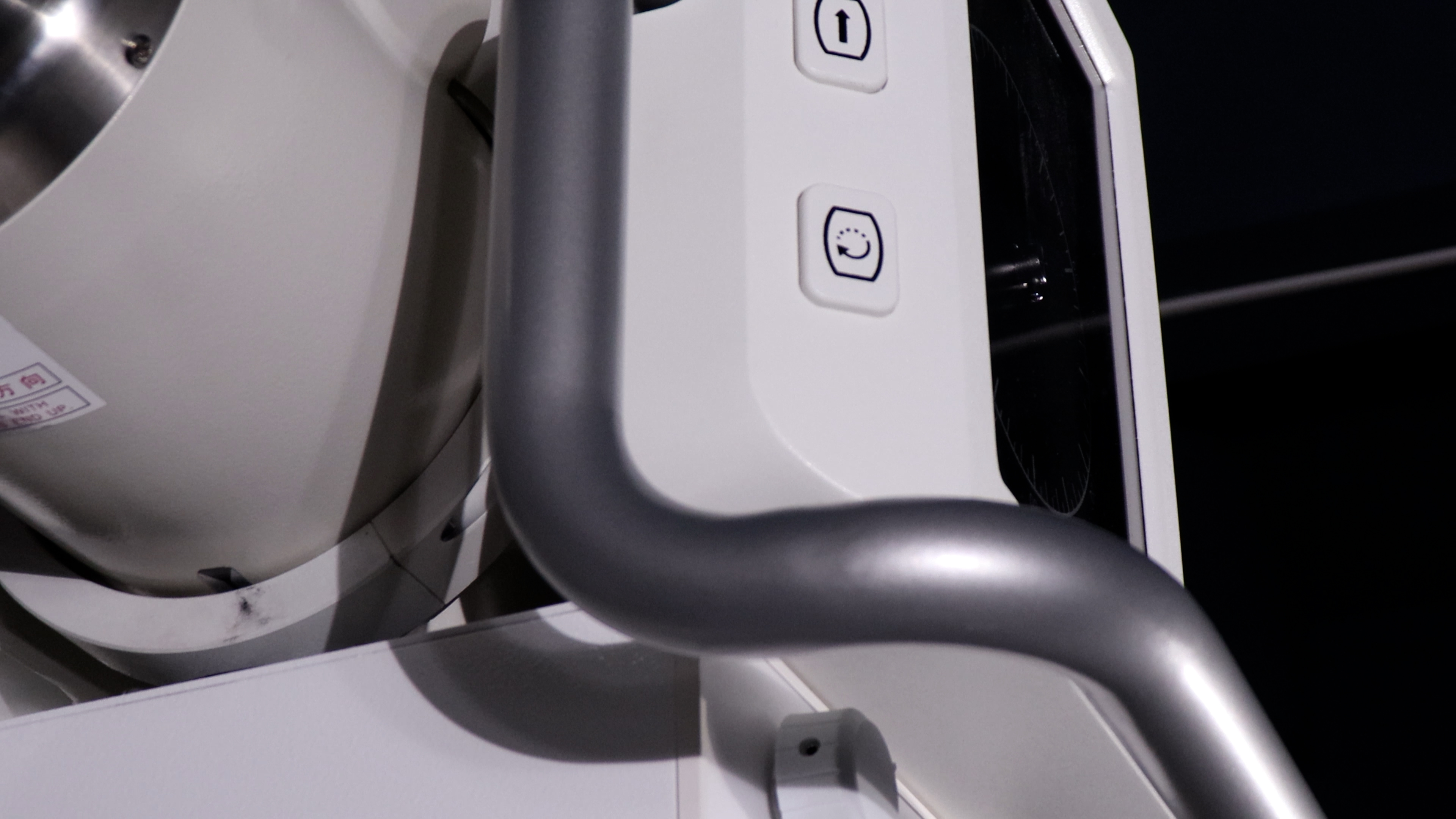
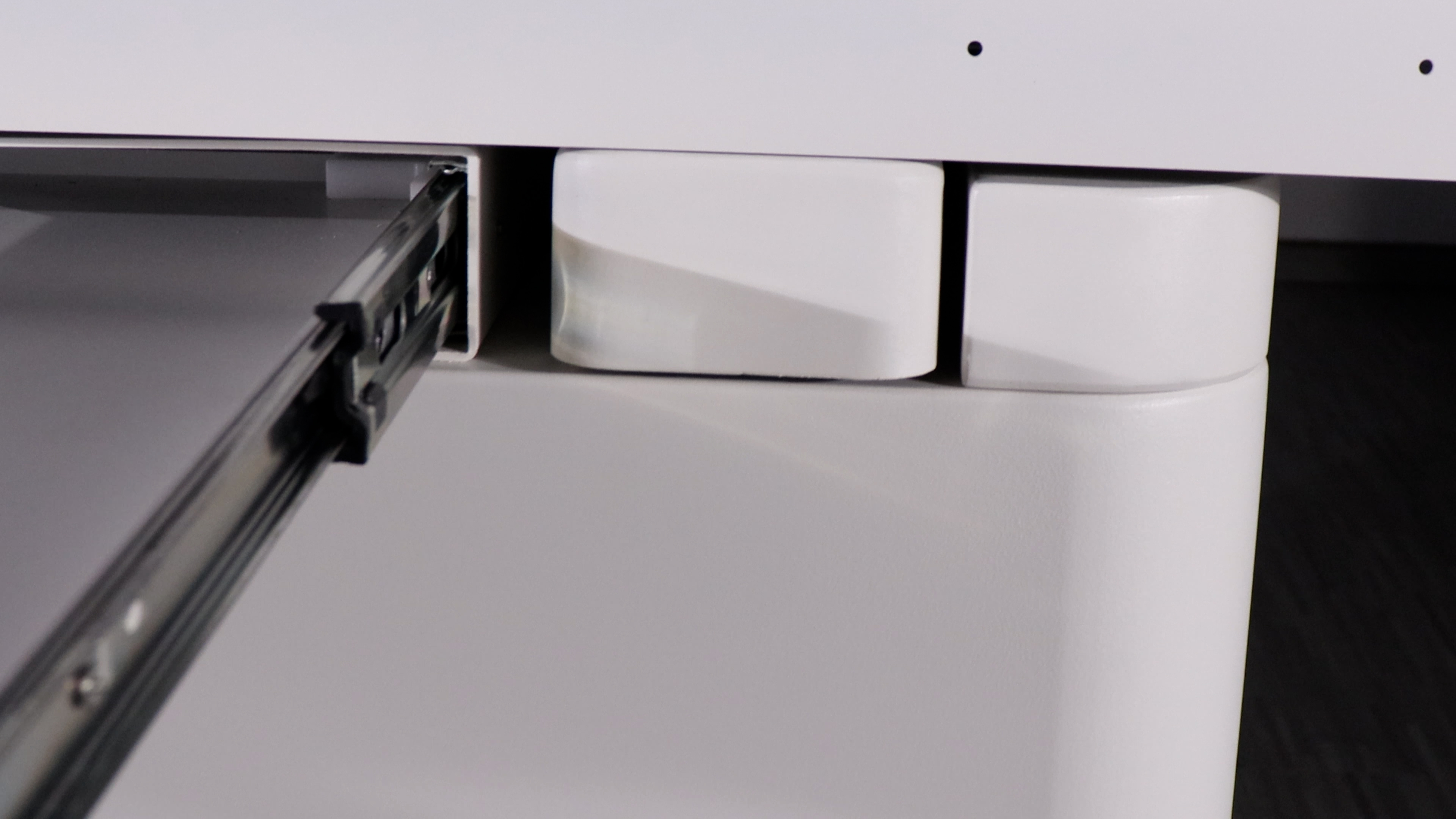
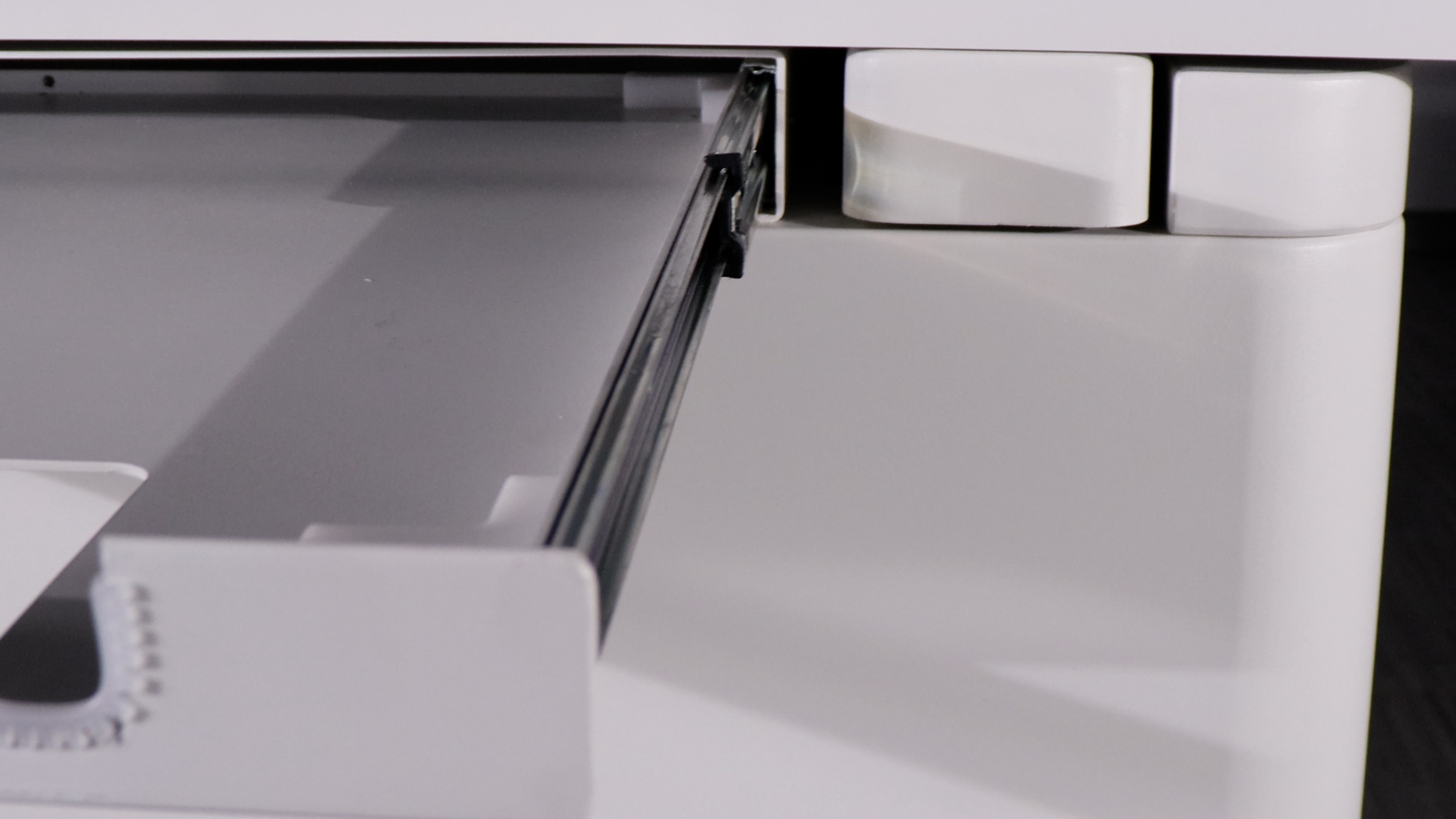
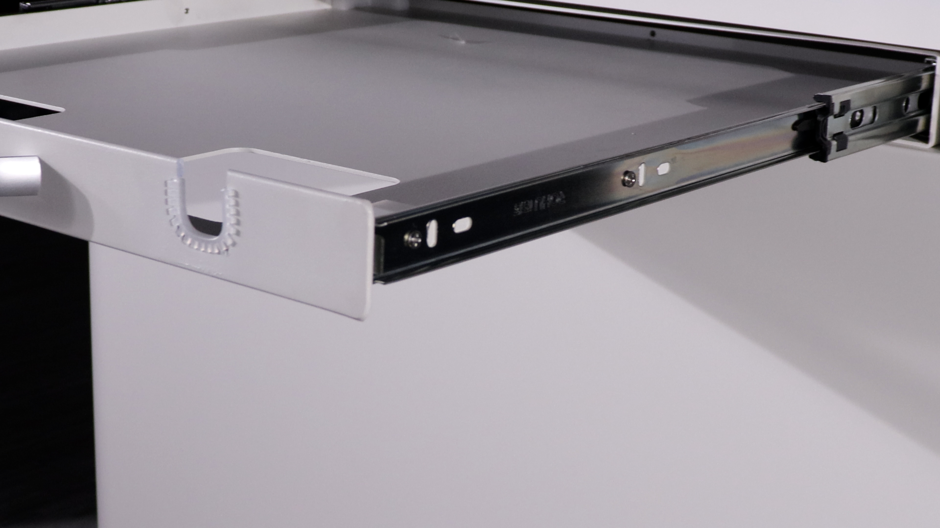
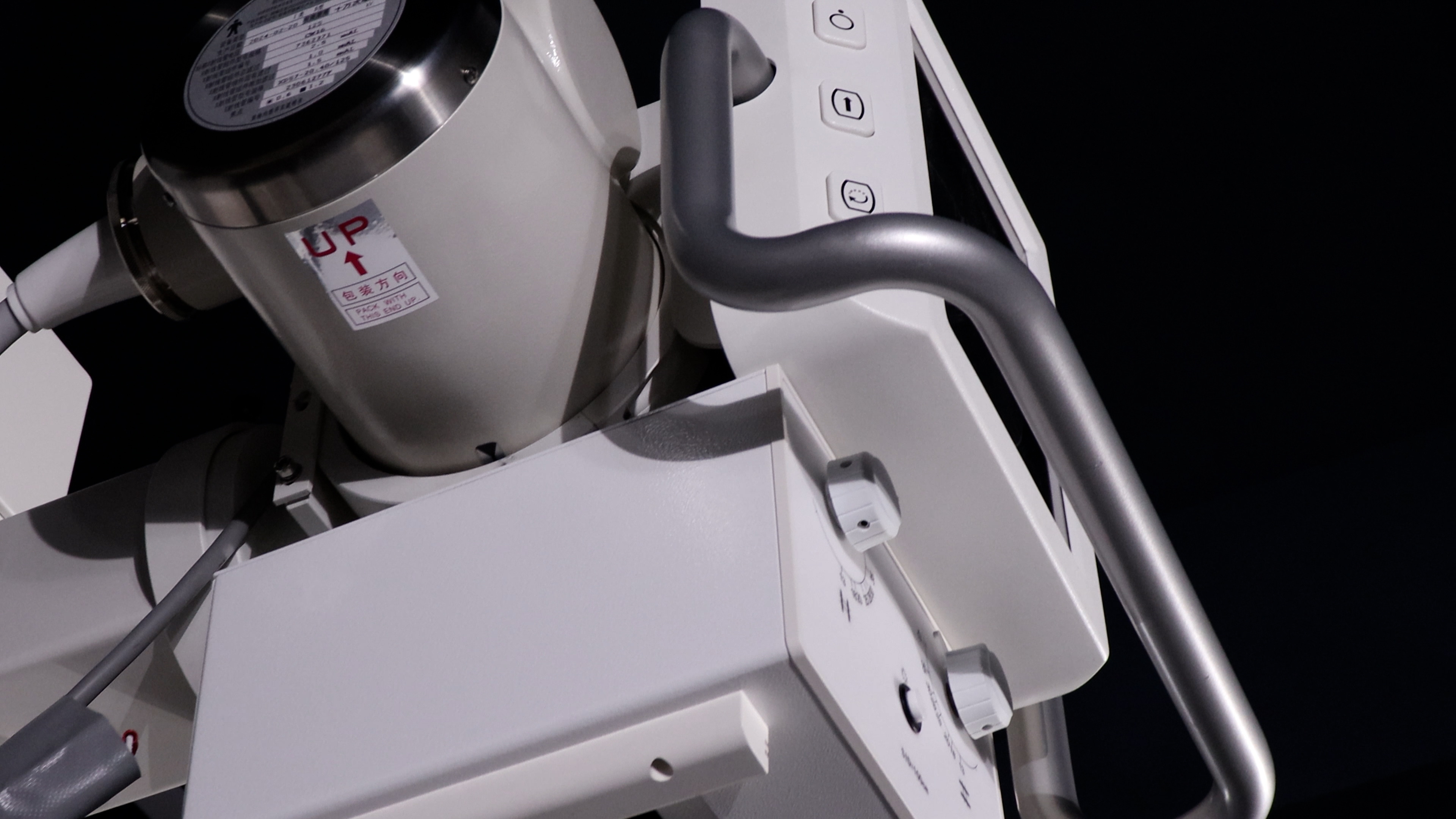
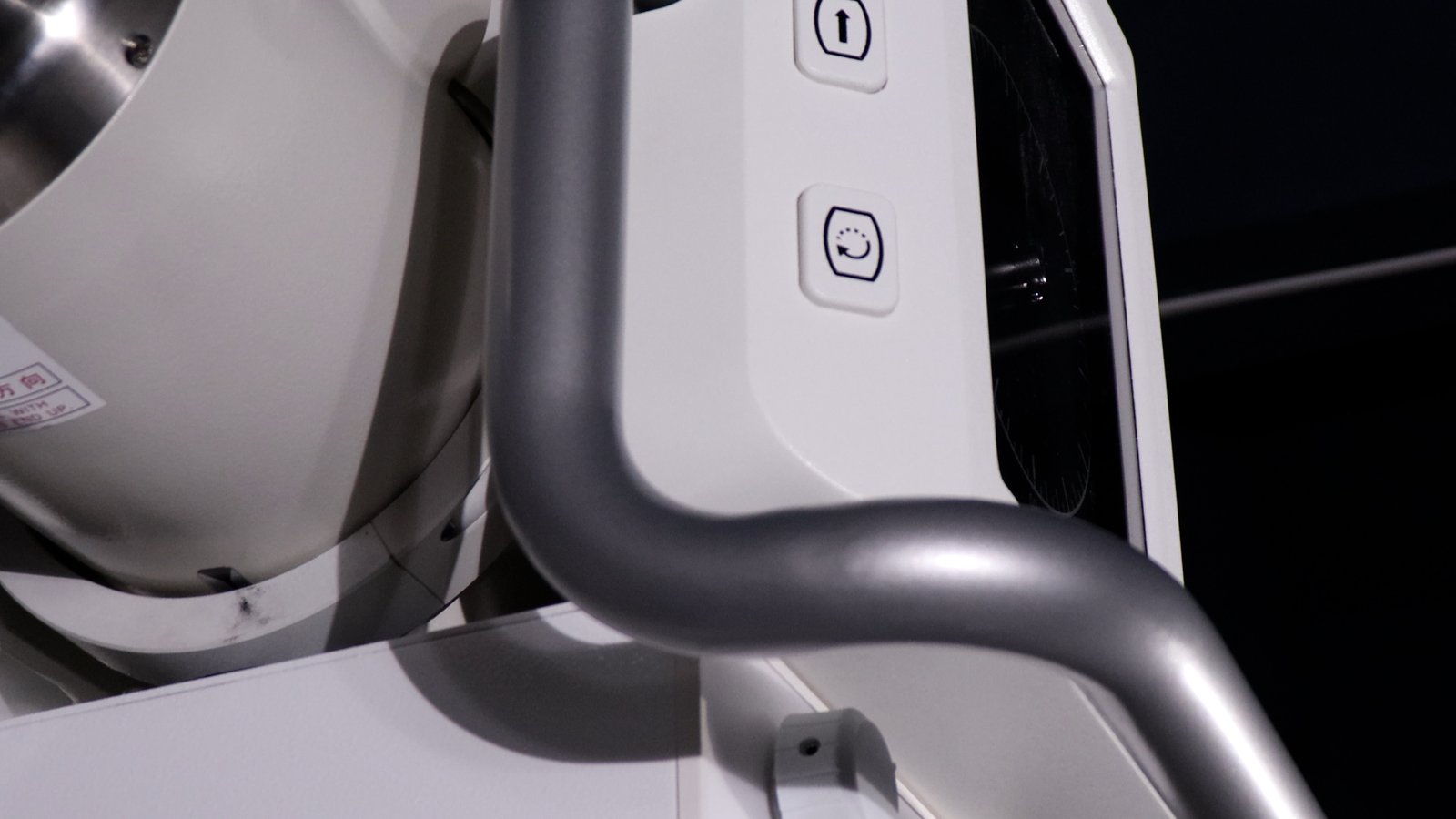
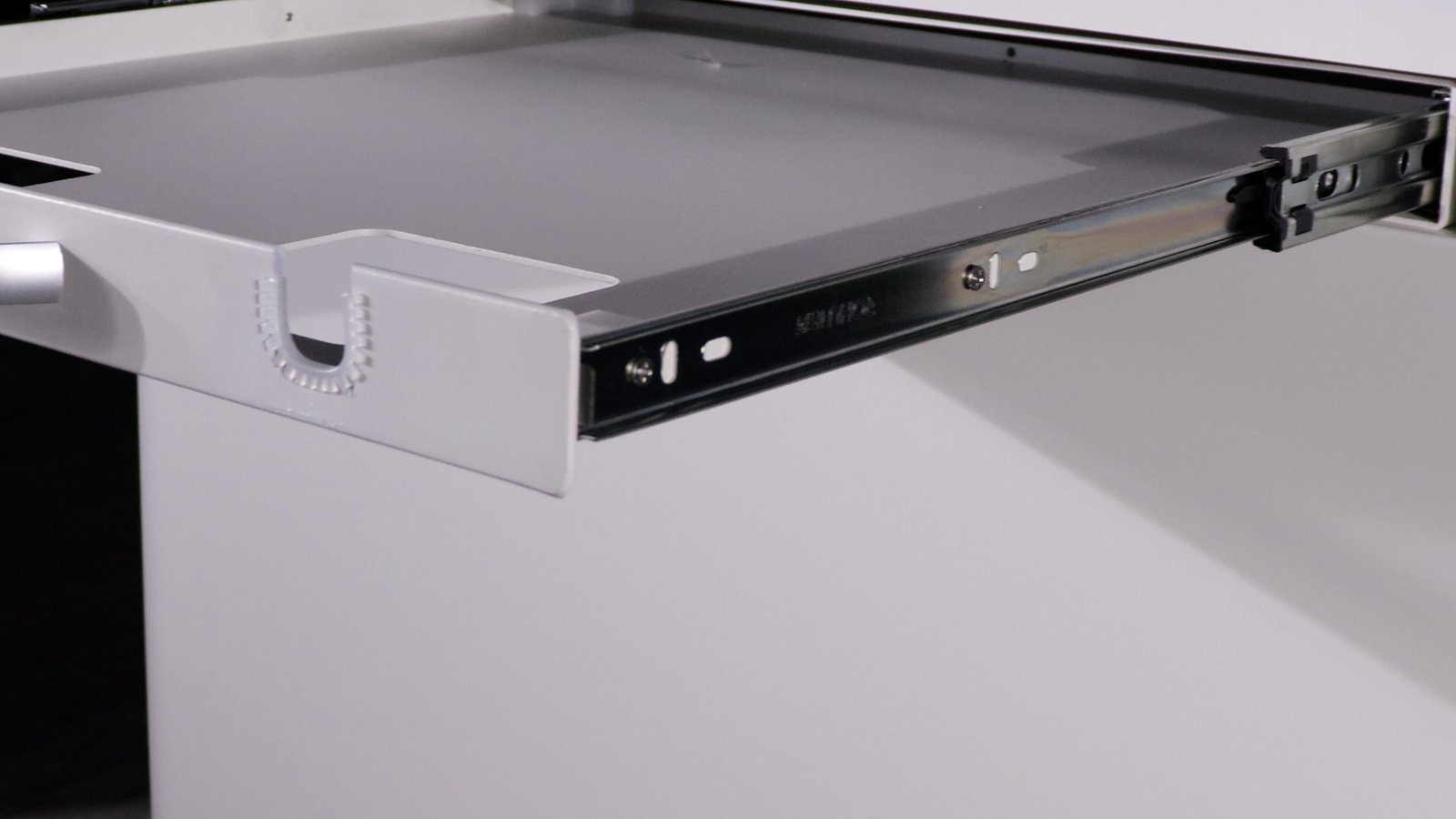
Detail views of the gantry column, rotating tube head and controls, four-way floating patient table, LED collimator, detector drawer rail, and acquisition workstation.
Platform Specifications (Pro Platform)
| High-voltage generator | 32 kW high-frequency, single phase 110/220 VAC (upgrade slot reserved for dynamic generator) |
| kV / mA range | 40 – 150 kV / 10 – 400 mA |
| Exposure time | 1 – 10000 ms |
| X-ray tube | Canon E7240X (max 125 kV, 0.6 / 1.2 mm focal spots, anode input 15 / 30 kW) |
| Gantry | Elevating, rotating column: adjustable SID / 90° tube rotation for horizontal beam / standing views |
| Positioning | Recumbent / standing / natural posture, single-operator workflow |
| Patient table | 1400 × 700 mm, phenolic resin, float X 75 ±10 mm / Y 200 ±10 mm |
| Collimator | LED light source, 1.0 mm Al filtration at 75 kV |
| Workstation | Industrial PC + 21.5″ touchscreen + 23.8″ monitor |
| Power supply | Single-phase mains, no three-phase supply required |
Flat-Panel Detector (iLume 4343 Standard)
| Detector type / scintillator | a-Si / CsI |
| Active area | 43 × 43 cm |
| Pixel matrix | 3072 × 3072 |
| Pixel pitch | 139 µm |
| A/D conversion | 16 bit |
| Min. detectable dose (RQA5) | 14 nGy |
| Spatial resolution | 3.6 LP/mm |
| Image acquisition | 1 s |
| Data interface | GigE |
| Sealing | IP65 |
Upgrade option: original iRay Venu 1717XV (43 × 43 cm / 2912 × 2912 / 139 µm / 3.6 LP/mm / 16 bit), serial number verifiable through iRay’s official channel.
Software Configuration
| Acquisition workstation | In-house software with Caravaggio tone engine and body-size automatic exposure |
| AI imaging assistant | Draft report generation; veterinarian reviews and signs off |
| Pro software package | AI lesion overlay display |
| DICOM | DICOM 3.0 — Store, Print, and Worklist |
| Cloud imaging | Cloud image storage and cross-device access supported |
Clinical Applications
- Advanced general practice: orthopaedic, thoracic and abdominal radiography, pre-surgical evaluation, and post-operative rechecks across all canine and feline body sizes
- Standing weight-bearing evaluation and natural-posture views (90° horizontal-beam projection)
- Fine orthopaedic detail at the 0.6 mm small focal spot
For single-exposure full-spine studies in giant-breed canines, a 17 × 24″ large-panel variant is available on request.
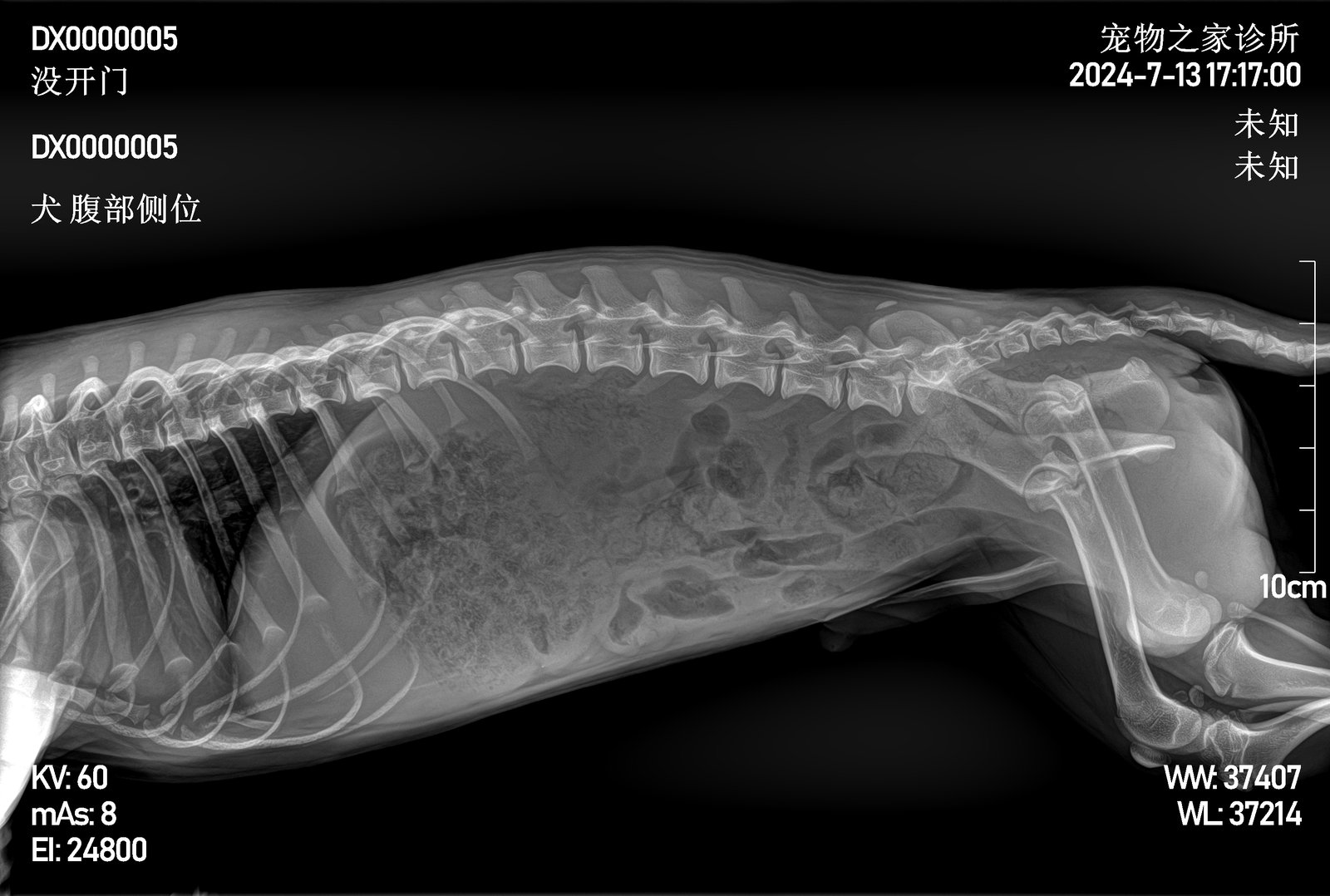
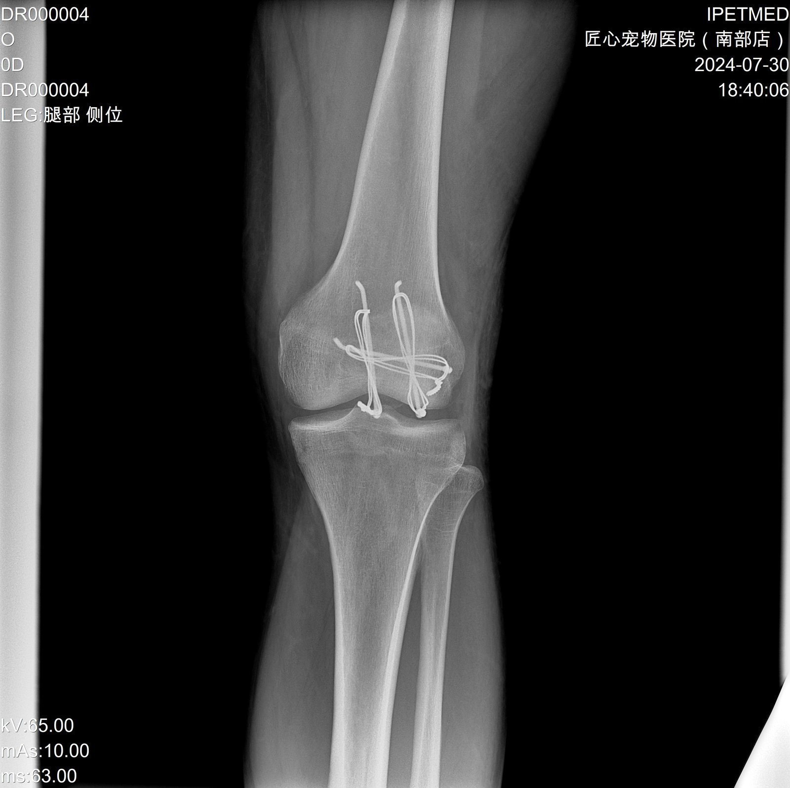
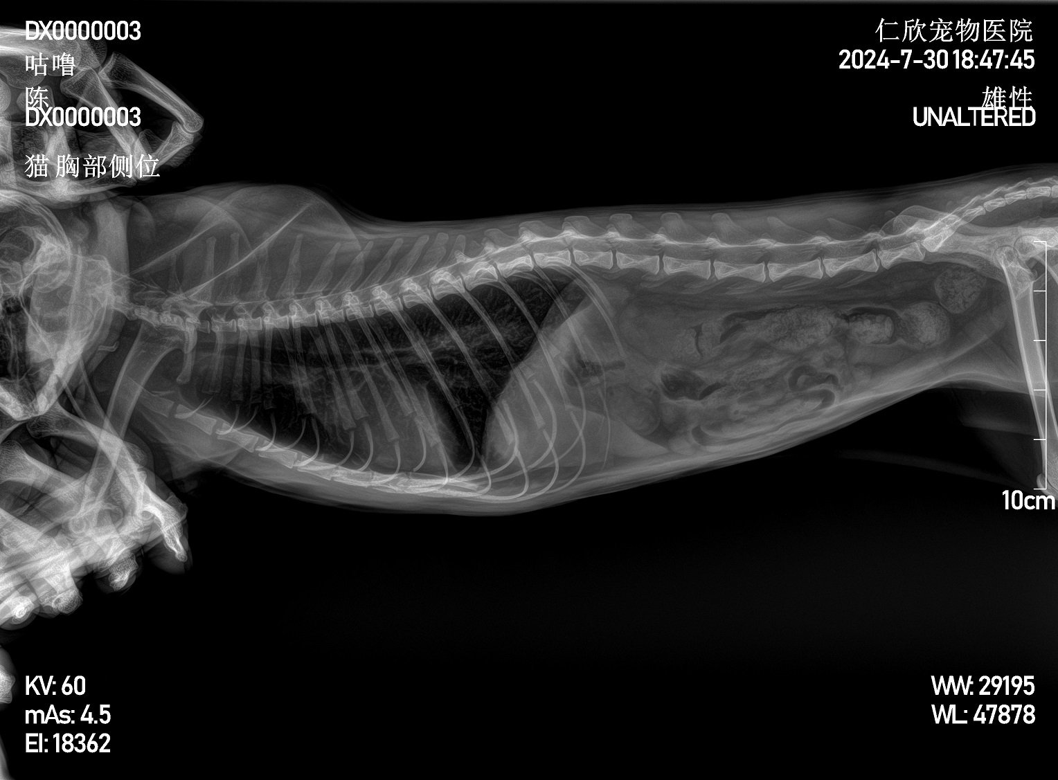
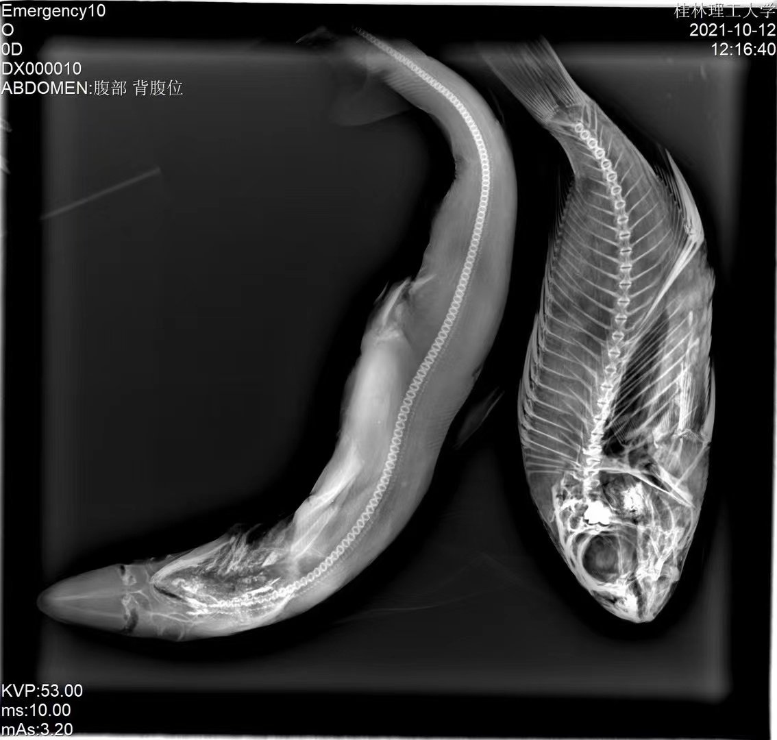
Clinical Imaging Samples
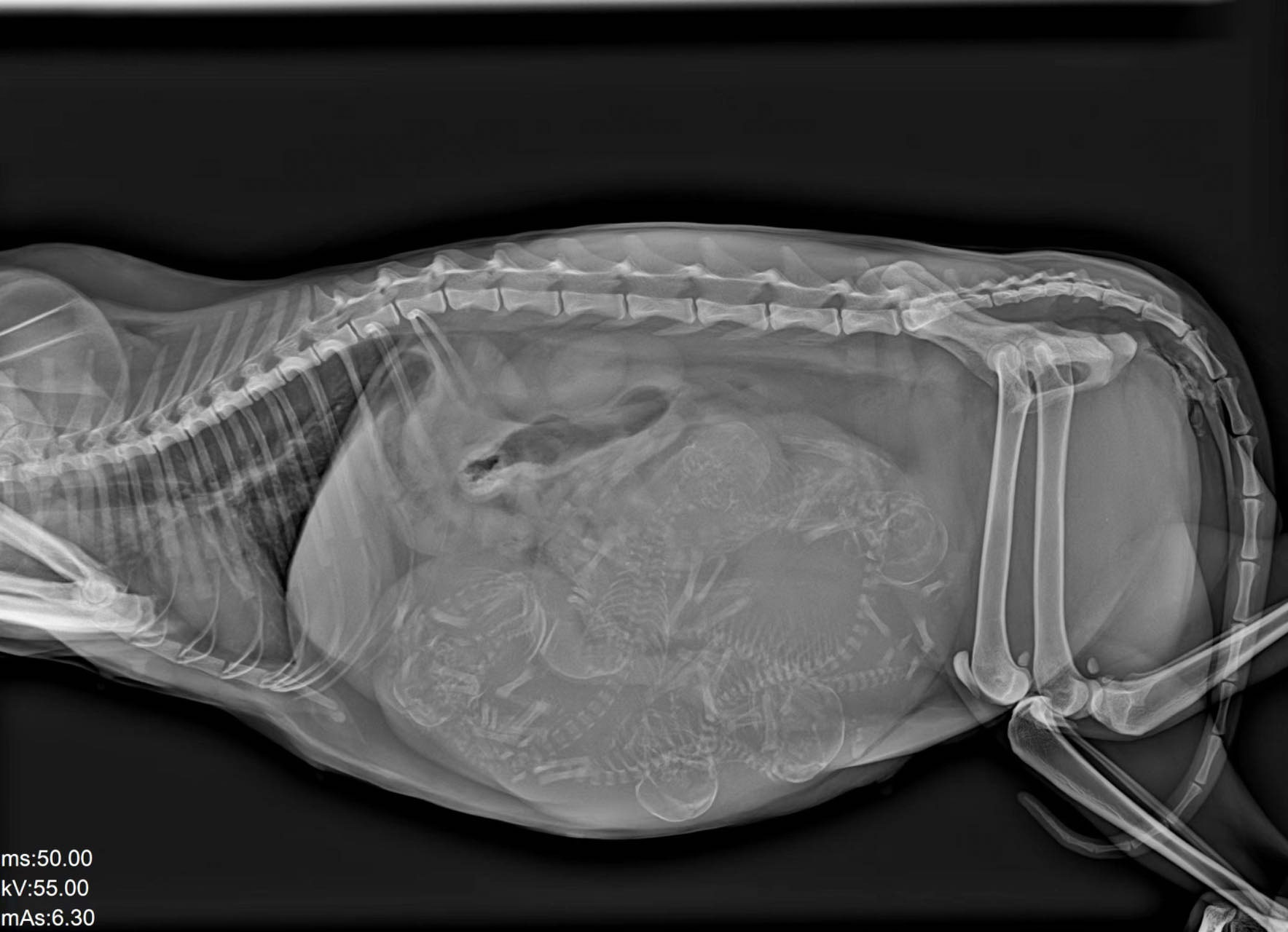
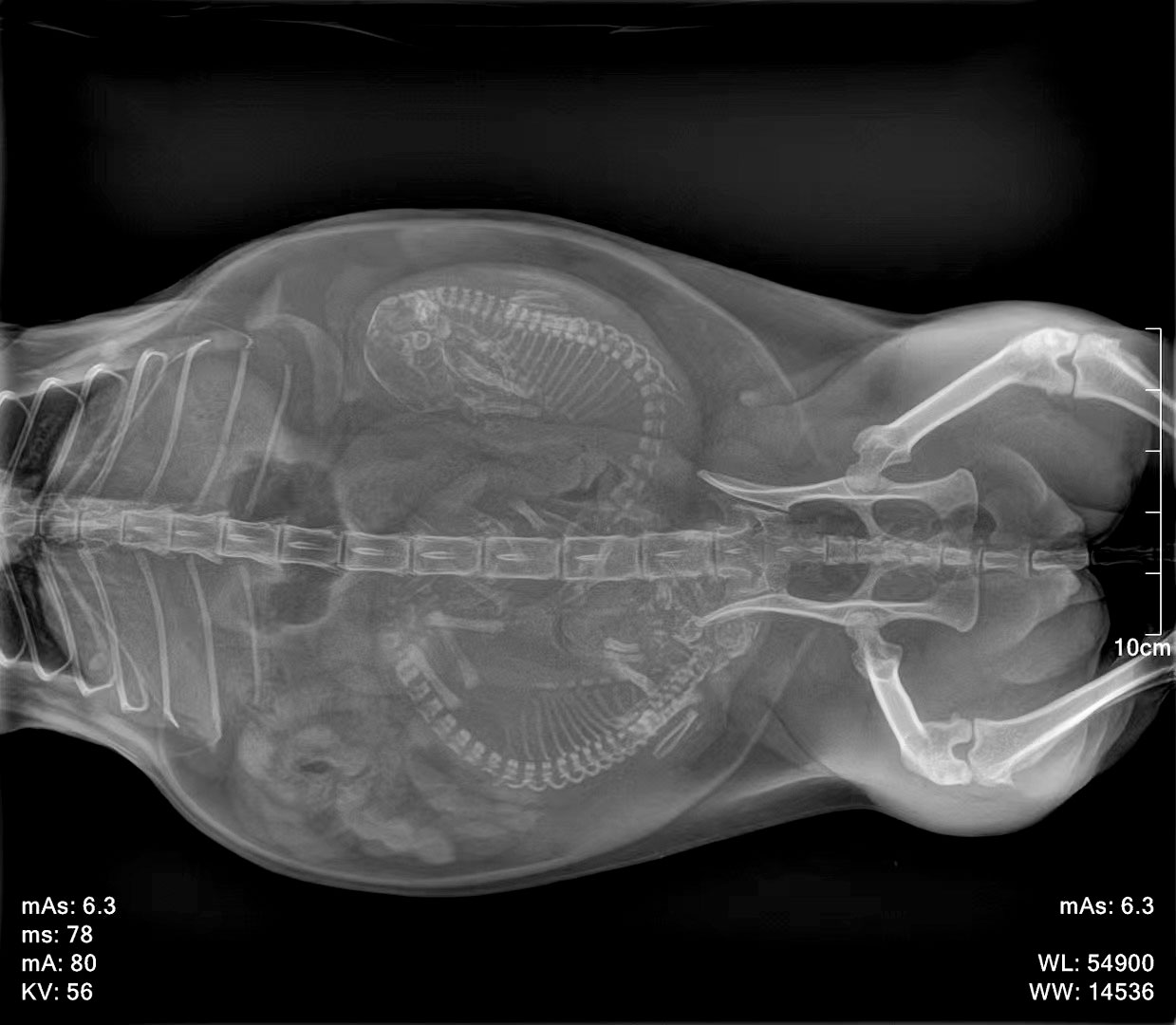
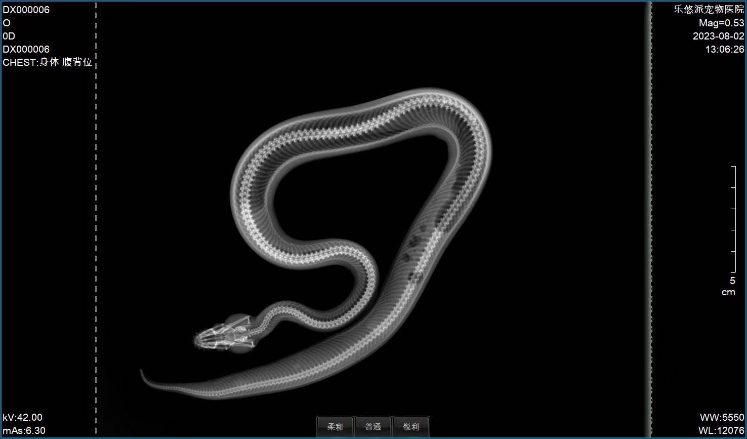
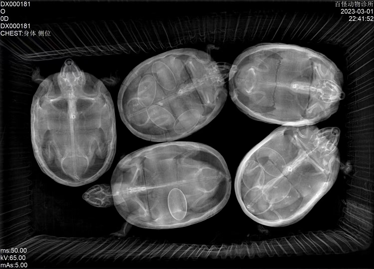
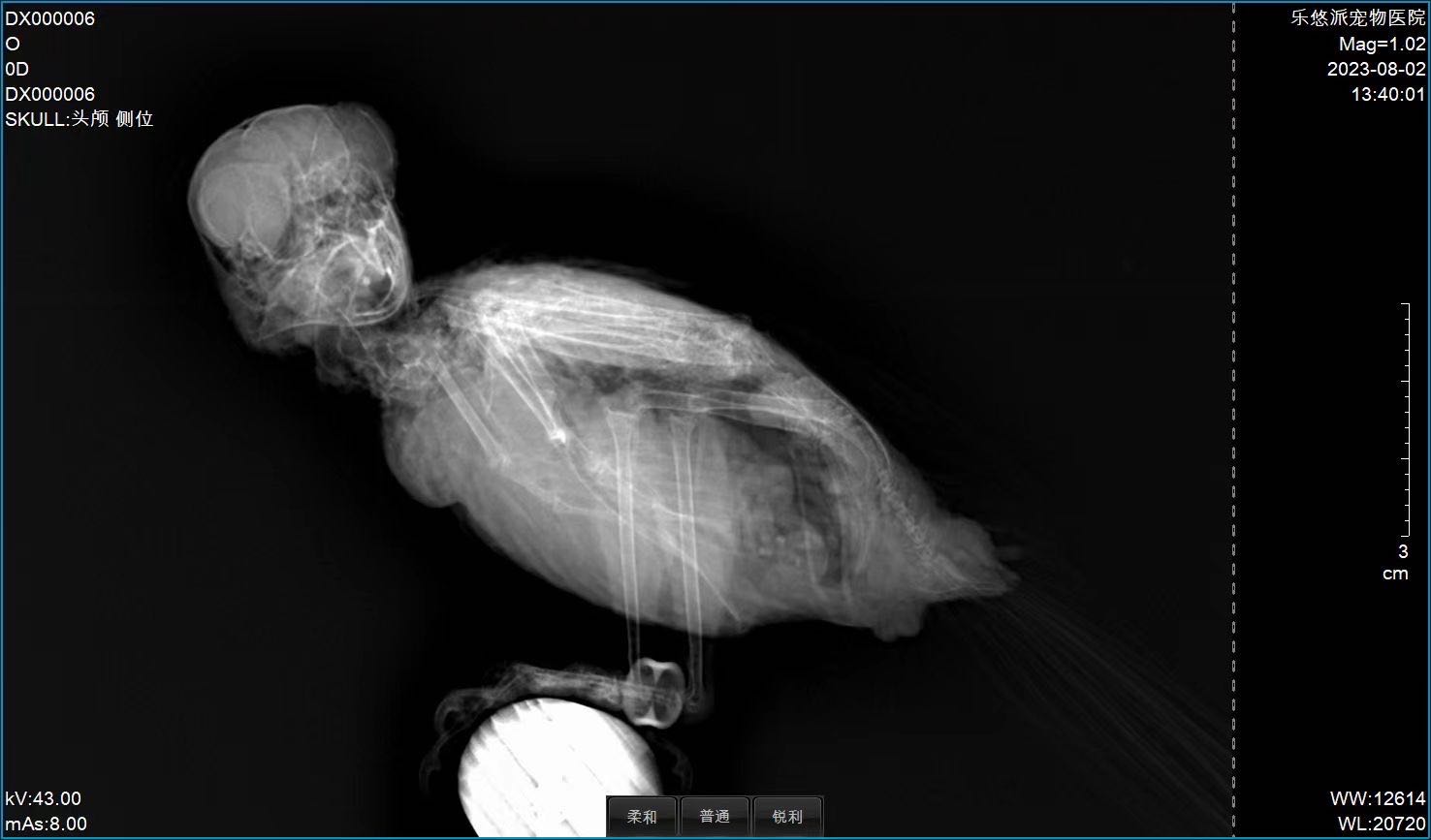
Representative radiographs from the iPet DR imaging chain across companion-animal and exotic species.
Workflow & Installation
Single-phase mains power. The elevating, rotating gantry lets the same room switch between tabletop and standing workflows without re-installation. Engineer-led installation, calibration, and operator training included.
iPet-400Pro installation walkthrough (English narration, ~4 min)
Package Contents
- 32 kW high-frequency generator + control box
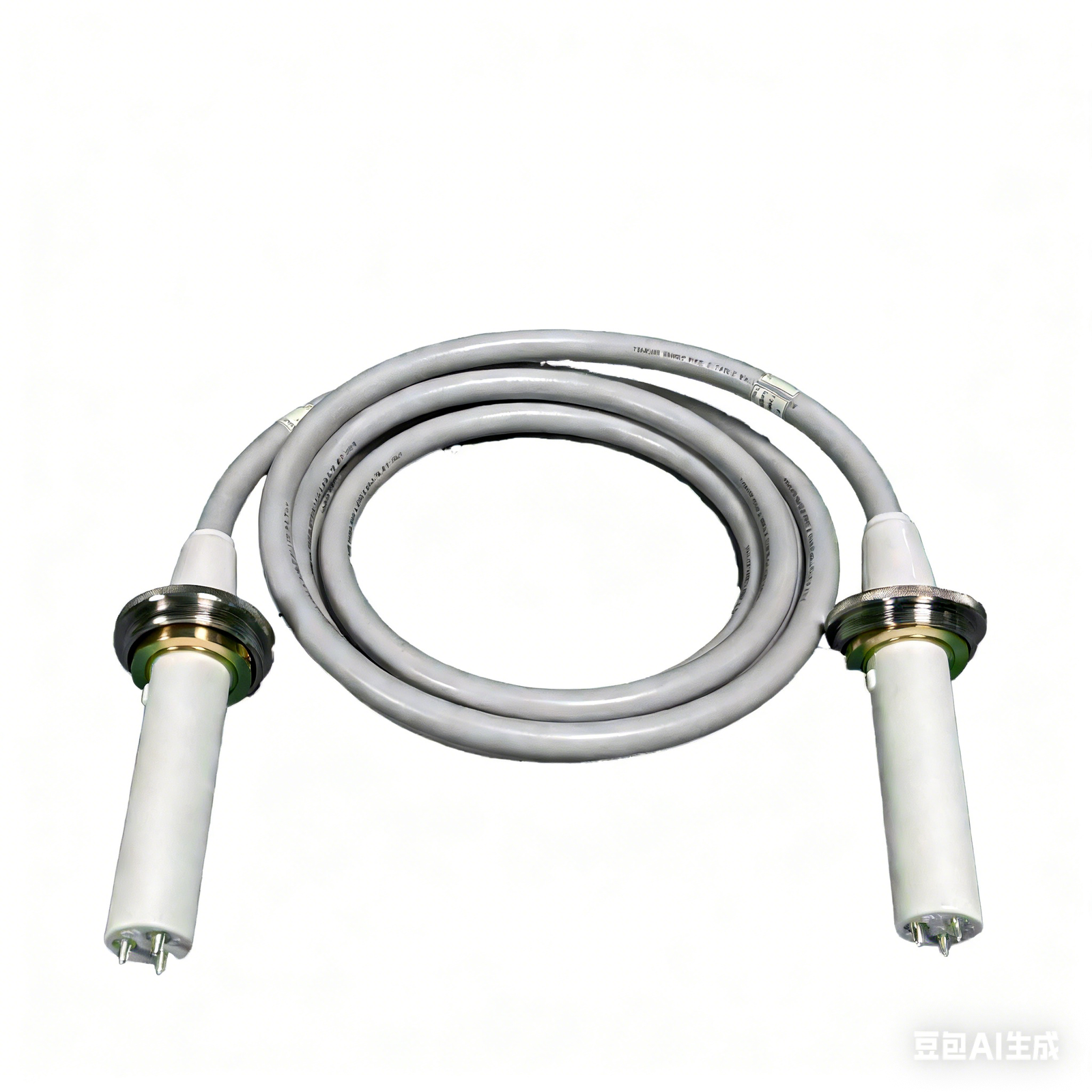
- X-ray tube assembly (Canon E7240X) + high-voltage cables
- iLume 4343 flat-panel detector (43 × 43 cm) + adapter
- LED collimator
- Elevating, rotating gantry + four-way floating patient table (1400 × 700 mm)
- Acquisition workstation: 23.8″ display, 21.5″ touchscreen, PC, software dongle
- V-shape positioning set (1 large + 1 small)
- Lead protection set: gown, hat, scarf, glasses, gloves
- Foot switch, remote control, network cable
Downloads
Why Choose iPet-400Pro
- Gantry freedom — where this price class typically buys a fixed table and fixed tube, the Pro invests in gantry articulation and an upgrade path.
- Built not to be outgrown — reserved upgrade slots for panel, tube, and generator protect the initial investment; static can become dynamic.
- Pro software package — AI report drafts and AI lesion overlay display included from day one.
Warranty & Support
Two-year warranty on hardware and software. Optional extended warranty available per year. Engineer-led on-site installation, calibration, and training included.
FAQ
Q: It shares a panel with iPet-400AI — what justifies the higher price?
The Pro is not priced on the panel. It is priced on the gantry and the upgrade rights: the elevating, rotating column completes recumbent, standing, and natural-posture views with one operator, and the panel, tube, and generator all carry reserved upgrade paths.
Q: Can it be upgraded to dynamic DR later?
Yes. The Pro platform reserves the upgrade path; a static system can be upgraded to a dynamic configuration without replacing the machine.
Q: What is standing-view imaging used for?
With the tube rotated 90° for horizontal-beam projection, the system supports standing weight-bearing evaluation and natural-posture views, reducing restraint difficulty.
Q: Can a giant-breed full spine be captured in one exposure?
The standard 43 × 43 cm panel covers routine views; for single-exposure full-spine or bilateral-hip studies, specify the 17 × 24″ large-panel variant at order.
Q: Does the system require three-phase power?
No. Standard single-phase mains is sufficient.

Reviews
There are no reviews yet.