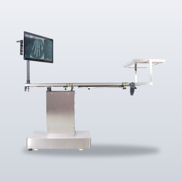
Mid-surgery, you need one look inside — iSurg-1 intra-operative imaging kit: spot radiography, not fluoroscopy.
Product Overview
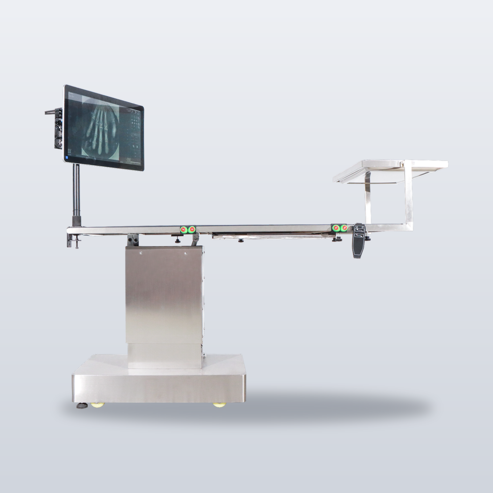
An intramedullary pin is placed — is it seated correctly? A foreign body is being retrieved — is the catheter where it should be? A retained root after extraction? These are the moments a surgeon most needs a radiograph — and moving the patient to the imaging room breaks sterility, extends anaesthesia, and takes two extra people. iSurg-1 brings the imaging to the table side: press once, image in one second, review, continue operating.
Spot imaging, not fluoroscopy — iSurg-1 takes static exposures only; it does not perform continuous real-time fluoroscopy.
Key Features
- Surgery continues, the patient stays put — images are taken at the table side; the sterile field stays intact and anaesthesia time is not extended.
- Dose lasts an instant, not the whole procedure — spot exposures emit for milliseconds and stop; cumulative staff dose is in a different class from continuous fluoroscopy.
- Most of the capability of conventional intra-operative imaging, at a fraction of the cost — no room conversion, no three-phase supply; standard mains and a corner of the theatre.
- Position it like a camera on a tripod — the tube head mounts on a wheeled stand that elevates and rotates to any angle: oblique, tangential, directly over the field.
- One kit covers three services — the intraoral sensor images single tooth roots; the 43 × 43 cm panel covers limbs and abdomen.
- Full-line software, not a bundled utility — in-house acquisition workstation, Caravaggio tone engine, AI report drafts (veterinarian sign-off), and DICOM worklist: the same suite as the flagship DR systems.
An Honest Comparison
| Conventional intra-operative fluoroscopy unit | iSurg-1 imaging kit |
|---|---|
| A major capital purchase | A fraction of the cost |
| Large footprint — usually a dedicated room | One camera-style stand + one panel |
| Projection angles limited by the C-arm geometry | Stand wheels, elevates, and rotates freely |
| Continuous fluoroscopy — high cumulative staff dose | Spot exposures, milliseconds of beam time |
| Dedicated room and electrical conversion required | Standard mains, wheel it in and use it |
| Real-time imaging during manipulation | No real-time fluoroscopy — static images only |
System Composition
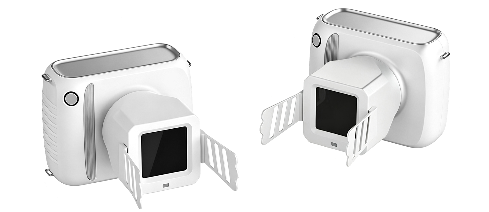
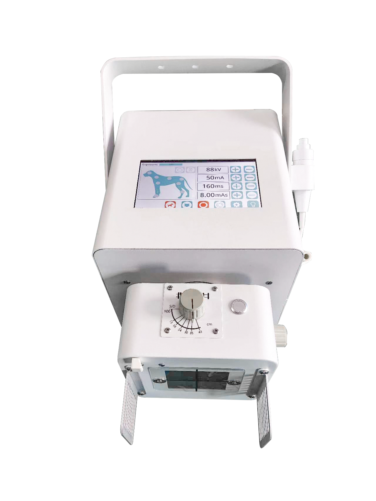
| X-ray tube head | iShot-70 handheld high-frequency head (integrated tube and generator, lithium-battery powered — no separate high-voltage cabinet) |
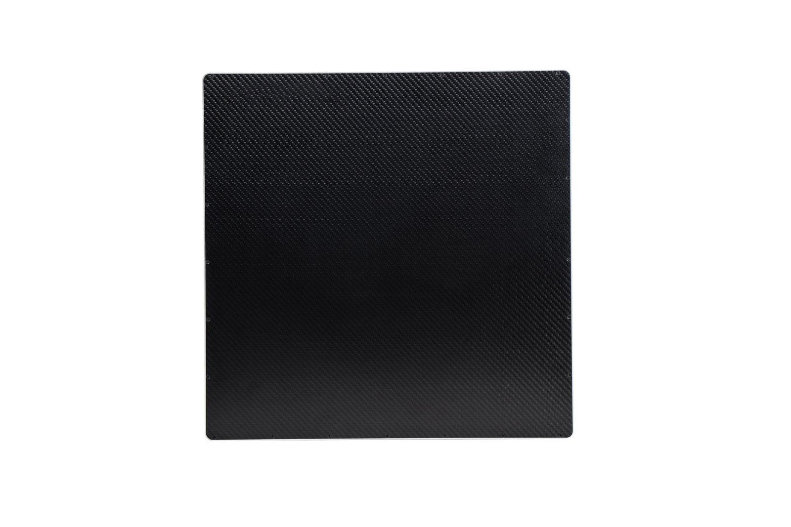
| Flat-panel detector | iLume 4343 panel (43 × 43 cm, wired, static) |
| Intraoral sensor | High-resolution dental intraoral sensor (DiD series, one model configured per order) |
| Mobile stand | Camera-style wheeled stand: mobile, height-adjustable, rotates to any angle |
| Workstation | Industrial PC + in-house acquisition software (customer-supplied PC also supported) |
X-ray Tube Head (iShot-70)
| Tube voltage | 70 kV |
| Tube current | 3 mA |
| Focal spot | 0.4 mm |
| Tube model | KL11-0.4-70 (anode target angle 12°) |
| Generator frequency | 80 kHz (high-frequency inverter) |
| Exposure time | 0.01 – 2 s |
| Duty cycle | 1:30 |
| Battery | 2500 mAh lithium; charger output DC 16.8 V |
| Weight | 1.9 kg (with battery) |
| Radiation leakage | <0.25 mGy/h |
Flat-Panel Detector (iLume 4343)
| Detector type / scintillator | a-Si / CsI |
| Active area | 43 × 43 cm |
| Pixel matrix | 3072 × 3072 |
| Pixel pitch | 139 µm |
| A/D conversion | 16 bit |
| Min. detectable dose (RQA5) | 14 nGy |
| Spatial resolution | 3.6 LP/mm |
| Image acquisition | 1 s |
| Data interface | GigE |
| Sealing | IP65 |
Intraoral Sensor (DiD Series — one model configured per order)
| Item | DiD2030 | DiD2636 | DiD2530 |
|---|---|---|---|
| Active area | 20 × 30 mm | 26 × 36 mm | 25 × 30 mm |
| Resolution | 800 × 1200 | 1300 × 1800 | 1242 × 1484 |
| Pixel size | 25 µm | 20 µm | 20 µm |
| Spatial resolution | 25 LP/mm | 25 LP/mm | 25 LP/mm |
| Scintillator | CsI | CsI | CsI |
| Interface / sealing | USB 2.0, IPX7 immersion-disinfectable | Same | Same |
| Exposure modes | Smart AED / preview mode | Same | Same |
Software Configuration
| Acquisition workstation | In-house software with Caravaggio tone engine and body-size automatic exposure |
| Intra-operative package | One-touch intra-operative capture + sterile-workflow interface |
| AI imaging assistant | Draft report generation; veterinarian reviews and signs off |
| DICOM / cloud | DICOM 3.0 Worklist support; cloud image storage supported |
Intended Use
- Dental: single tooth roots, periodontal assessment, intra-operative retained-root confirmation
- Orthopaedics: distal-limb fracture reduction and implant-position confirmation
- Abdomen: intra-operative foreign-body localisation and catheter-position confirmation in small and medium patients (recommended under 15 kg)
Continuous real-time fluoroscopy is not supported. For thick anatomy — large-breed canine thorax, spine, pelvis — use the iPet-400AI or a higher-tier system.
Operating Requirements & Installation
| Power supply | Standard single-phase mains — no three-phase supply |
| Room requirements | No dedicated radiography-room conversion; radiation shielding per local regulations (lead apparel supplied) |
Imaging Samples
First clinical sample images are being compiled; contact IWA for reference radiographs.
Package Contents
- X-ray tube head (integrated tube and generator, with lithium battery and charger)
- iLume 4343 flat-panel detector (43 × 43 cm) + cabling
- Dental intraoral sensor (DiD series, one model per order)
- Camera-style mobile stand (wheeled / height-adjustable / rotating)
- Acquisition workstation with in-house software (customer-supplied PC also supported)
- Lead protection set
Entry-Level Companion
Dentistry only? The IWA dental DR kit (tube head + intraoral sensor) covers exotic, canine, and feline oral imaging; adding the iLume 4343 panel and mobile stand later upgrades it to the full intra-operative kit.
Downloads
Why Choose iSurg-1
- Workflow efficiency — one press, one second to image; no broken sterility, no extended anaesthesia, no patient transfer.
- Dose control — spot exposures emit for milliseconds; cumulative staff dose is in a different class from continuous fluoroscopy.
- Practice economics — most of what conventional intra-operative imaging delivers, without the room, the electrical conversion, or the capital outlay.
Warranty & Support
Two-year warranty on hardware and software. Engineer-led installation and operator training included.
FAQ
Q: Is iSurg-1 a fluoroscopy unit?
No. iSurg-1 takes intra-operative spot exposures — static images only, no real-time fluoroscopy. For continuous dynamic imaging, see the iSmart-32Pro dynamic DR system.
Q: Which surgical scenarios does it cover?
Dental (retained-root confirmation), orthopaedics (distal-limb reduction and fixation checks), and small-to-medium-patient abdominal work (foreign-body and catheter localisation). For large-breed thorax, spine, or pelvis, use iPet-400AI or above.
Q: Does the theatre need conversion?
No room conversion is required. Standard single-phase mains is sufficient; radiation shielding follows local regulations, and lead apparel is supplied.
Q: How much dose does the surgical team receive?
Spot exposures emit only during the millisecond exposure window — cumulative dose is in a different class from continuous fluoroscopy. Protective apparel must still be worn per protocol.
Q: Is the software the same as the full-size systems?
Yes. The in-house acquisition workstation, Caravaggio tone engine, and AI imaging assistant (draft reports, veterinarian sign-off) are the same software suite as the flagship DR line.

Reviews
There are no reviews yet.