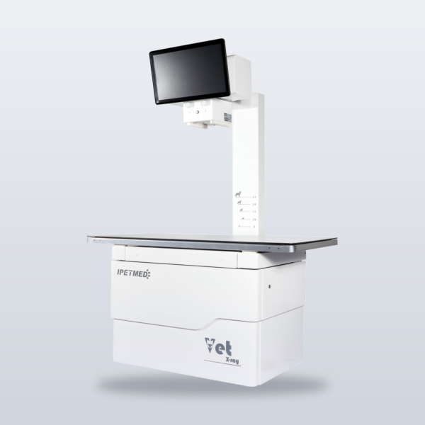
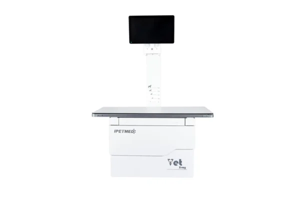
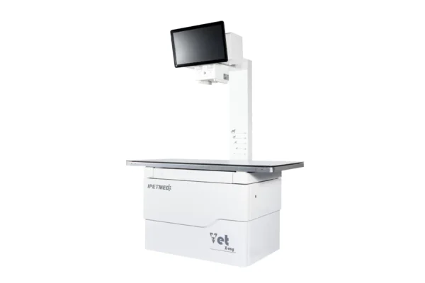
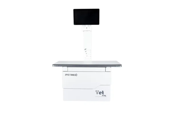
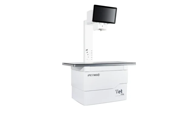
Sharp images that read themselves into a draft report — iPet-400AI, the AI-assisted DR workhorse for general small-animal practice.
Product Overview
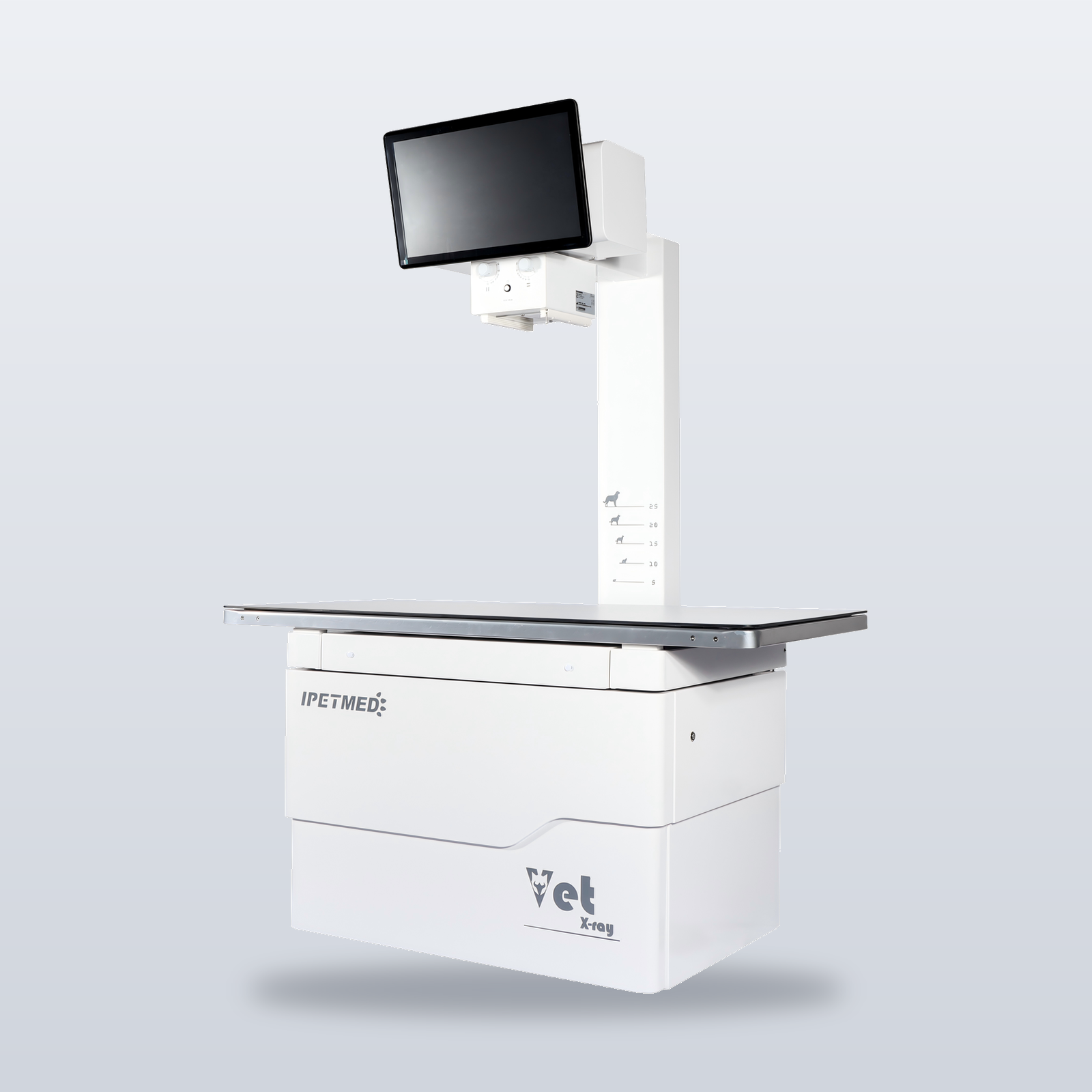
A general practice may run twenty exposures a day — orthopaedic, thoracic, abdominal, pre-anaesthetic, recheck. iPet-400AI is built for exactly that cadence: a 32 kW high-frequency generator and Canon E7239X rotating-anode tube keep output consistent through the day; the acquisition workstation sets exposure parameters automatically by body size, so retake rates stay low even with new staff at the console. After every exposure, the AI imaging assistant generates a draft report for the veterinarian to review, edit, and sign off — cutting reading time on routine studies.
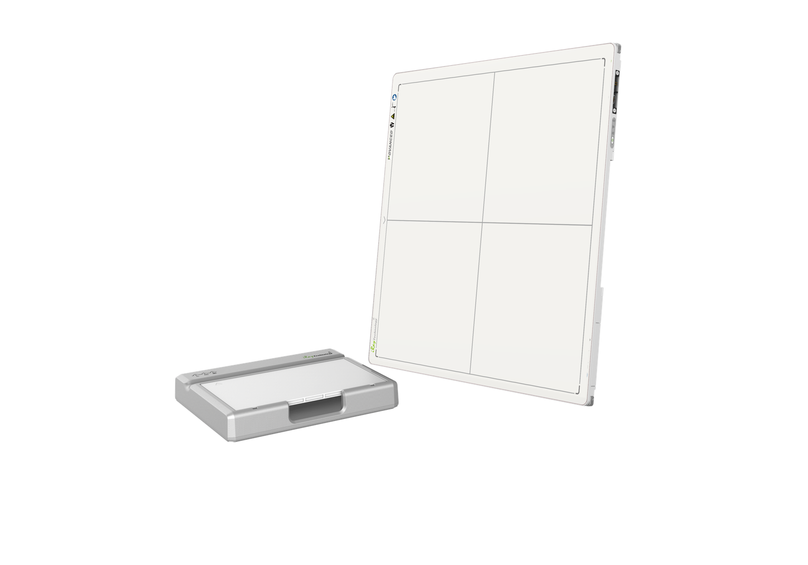
The iLume 4343 flat panel is IWA’s own-brand detector, manufactured by a specialist panel factory and tuned at SDK level with the in-house acquisition software — hardware and software calibrated as one imaging chain.
Key Features
- AI-assisted report drafts as standard — the AI assistant drafts, the veterinarian reviews and signs off; an assist tool, not a diagnostic replacement.
- Intelligent acquisition workstation — body-size automatic exposure, collimator auto-detection, and VHS cardiac measurement built in.
- iLume 4343 own-brand flat panel — 43 × 43 cm CsI detector with SDK-level tuning against the acquisition software.
- DICOM worklist — order straight to DR — exam orders created in practice-management software arrive at the DR workstation automatically; no double data entry, no patient mix-ups.
- Cloud image storage supported — images stay accessible when workstations change or a branch clinic opens.
- Manufactured in IWA’s own factory, on the same supply chain as an installed base of more than 5,000 systems.
How iPet-400AI Compares
| Common practice at this price point | iPet-400AI |
|---|---|
| Bare system + third-party acquisition software | Fully in-house software — one manufacturer accountable for machine and software |
| AI features priced as extras, or absent | AI imaging assistant included as standard |
| Unbranded detectors of unclear origin | Honestly labelled own-brand iLume panel, tuned to the imaging chain |
Product Gallery
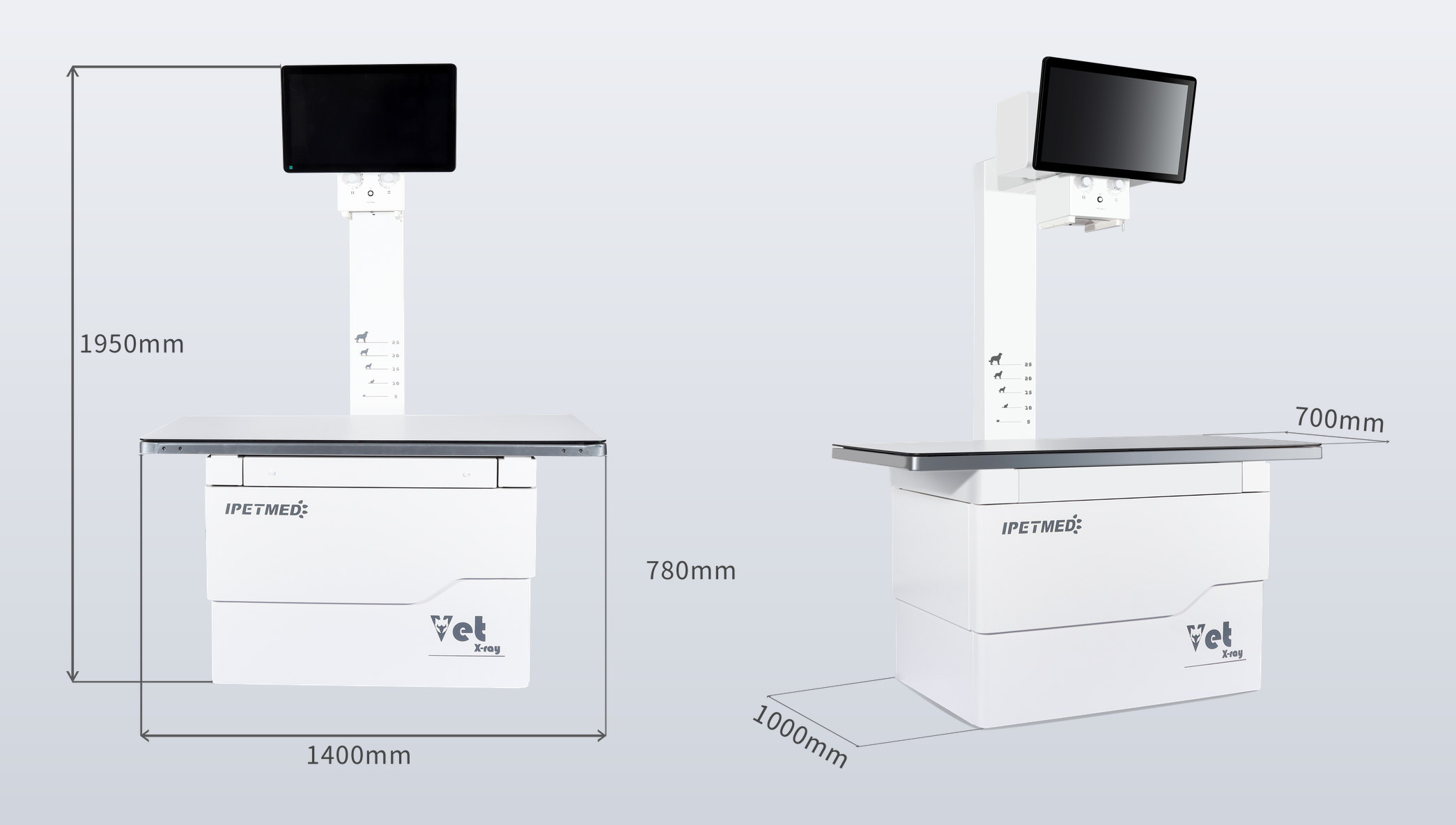
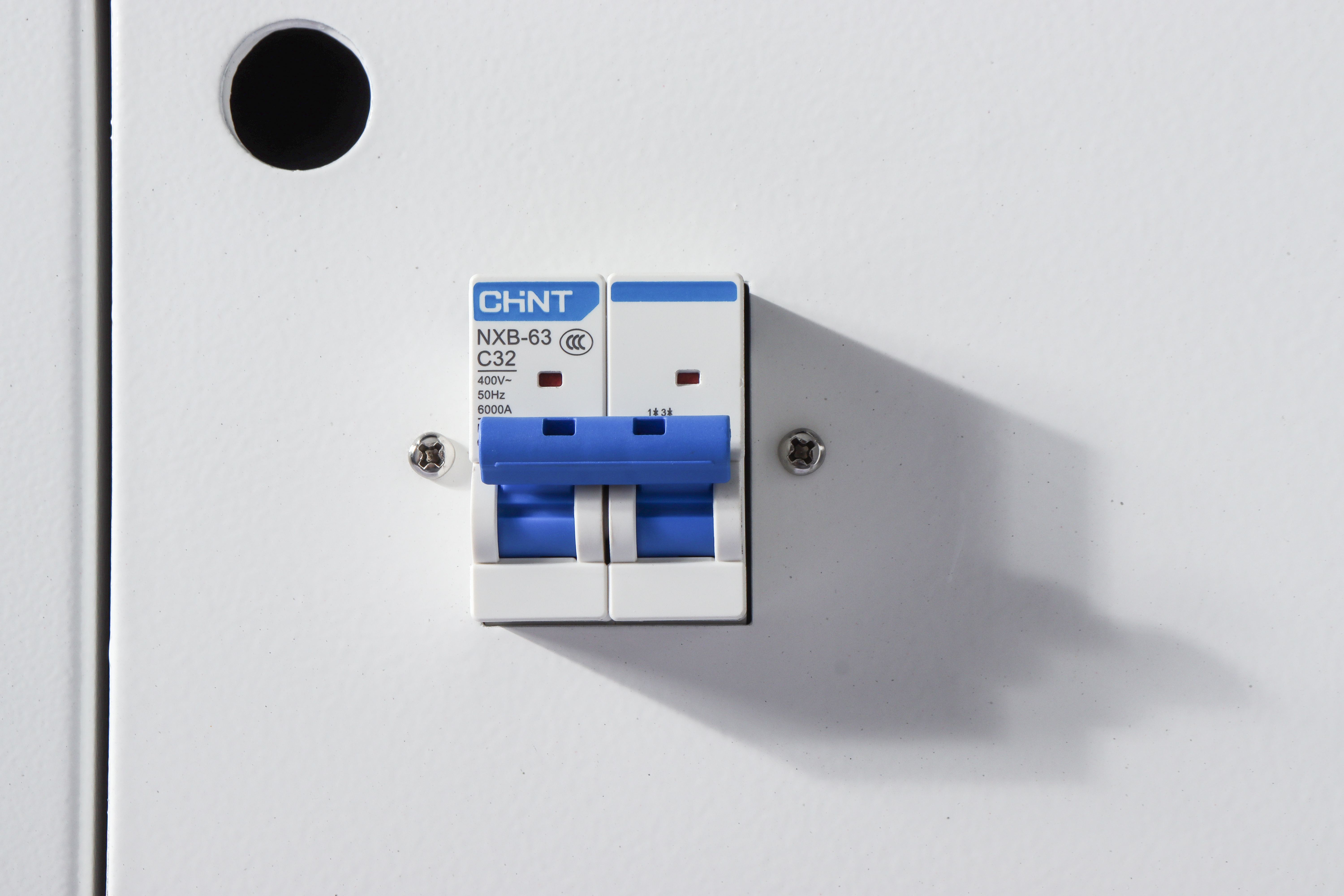
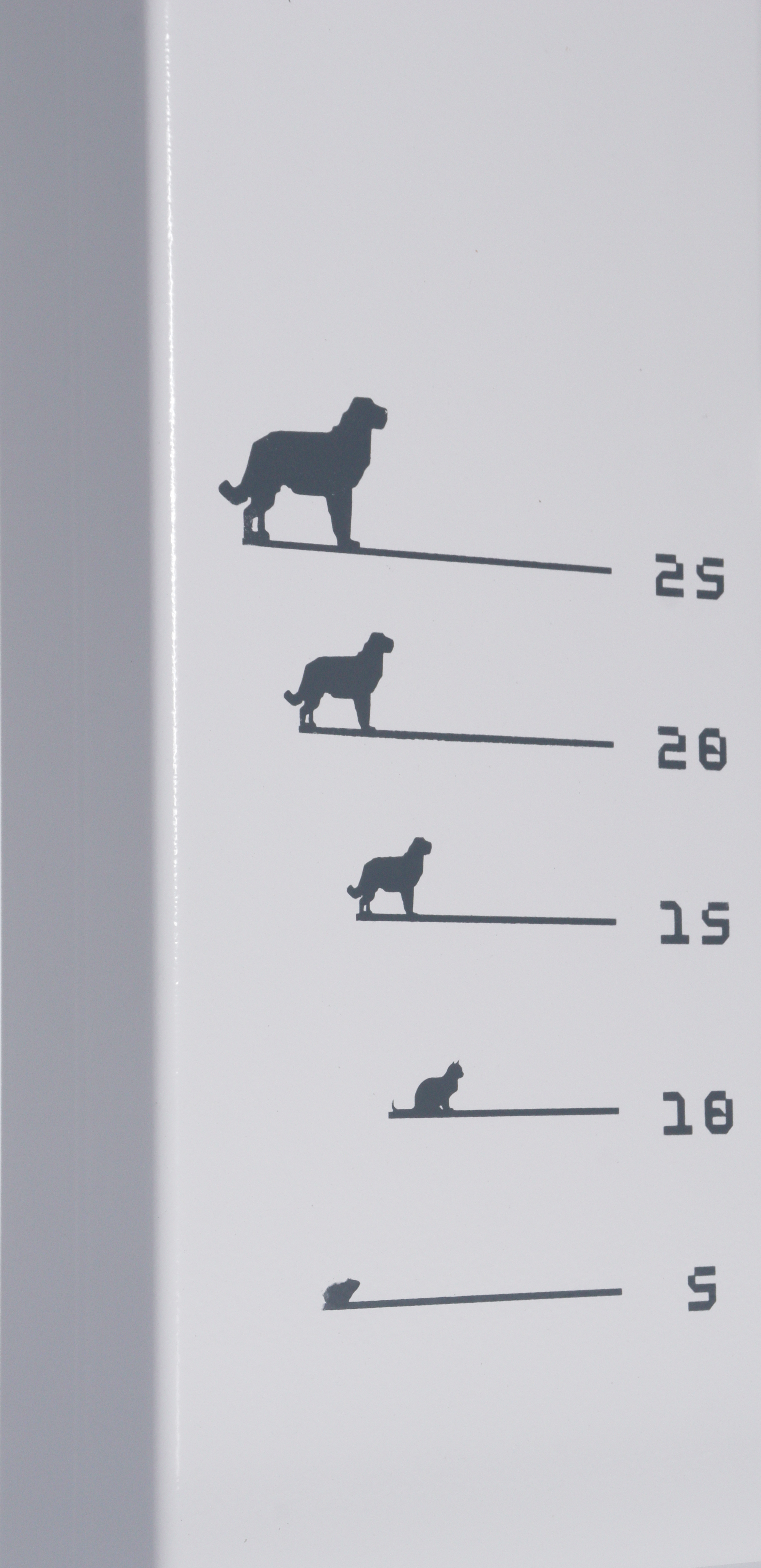
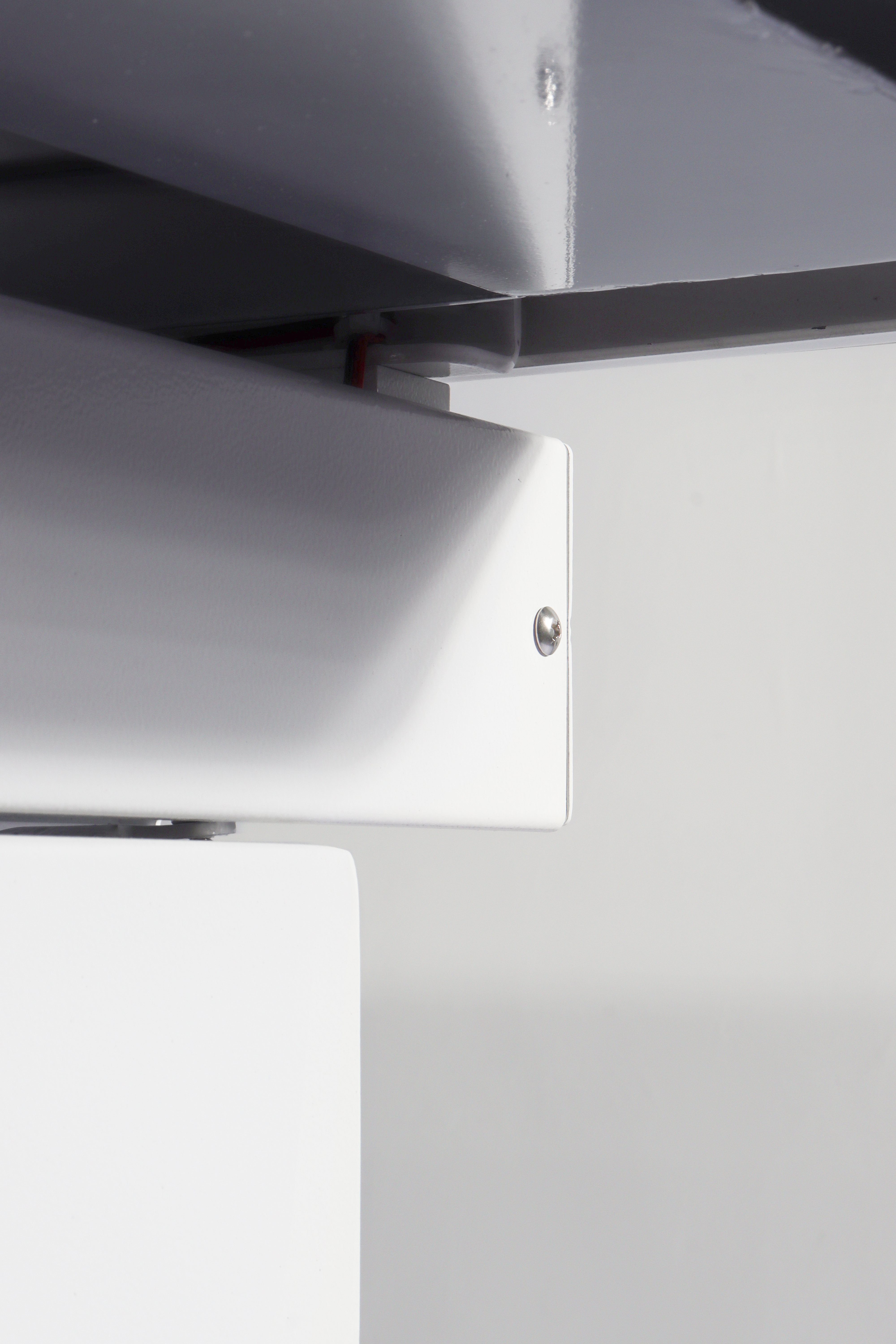
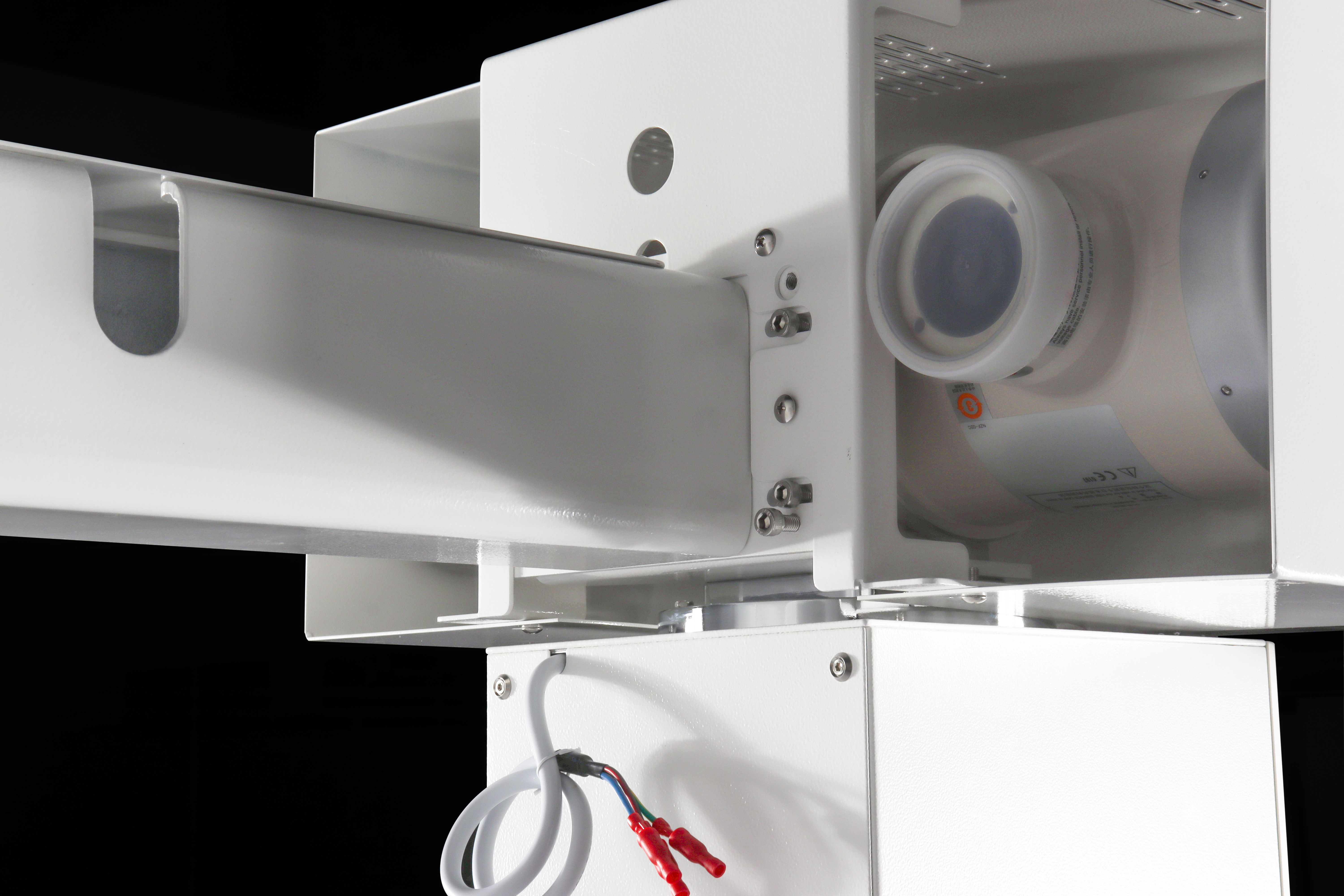
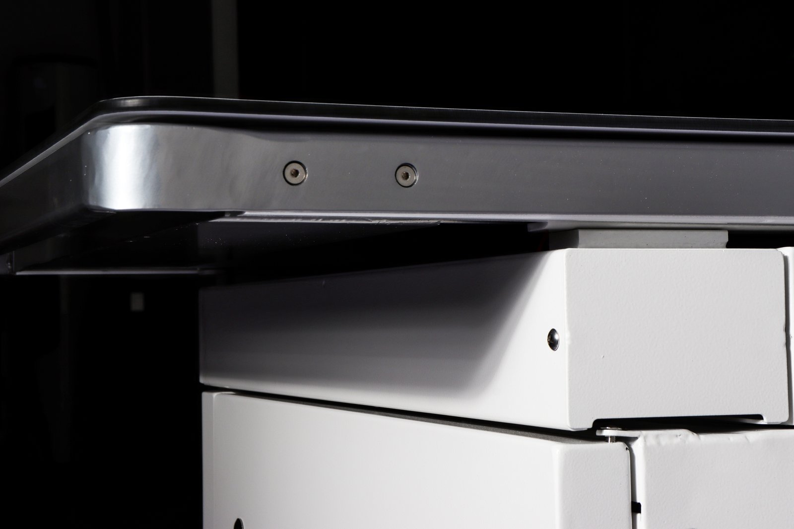
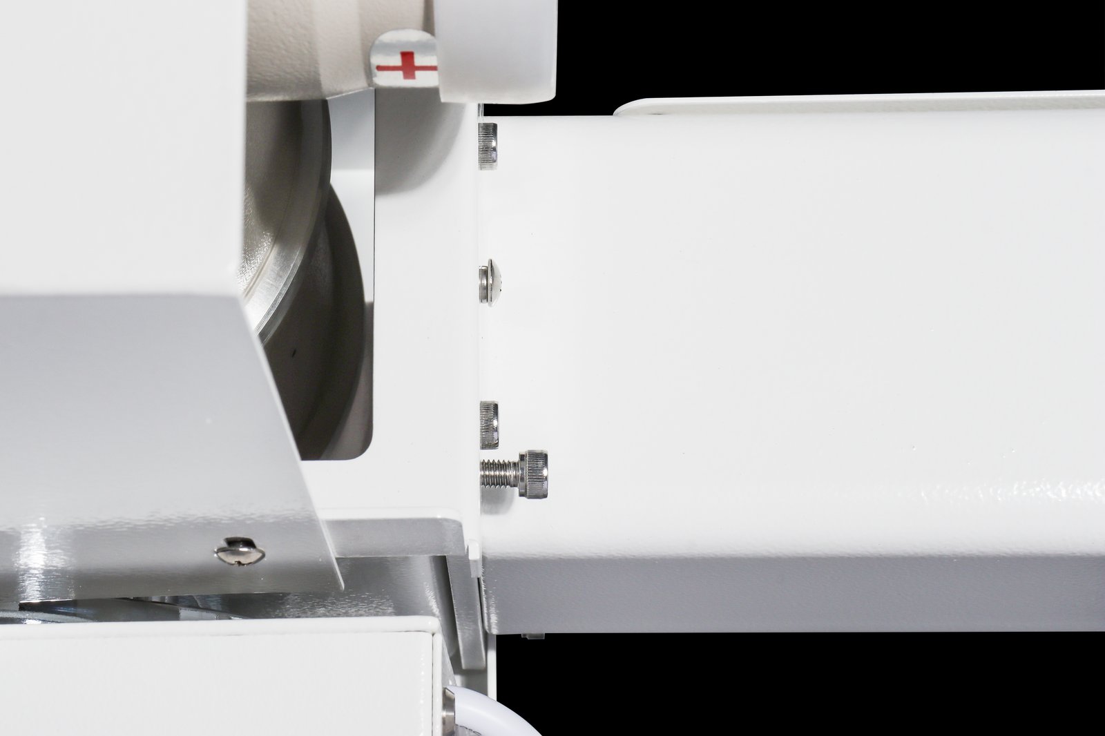
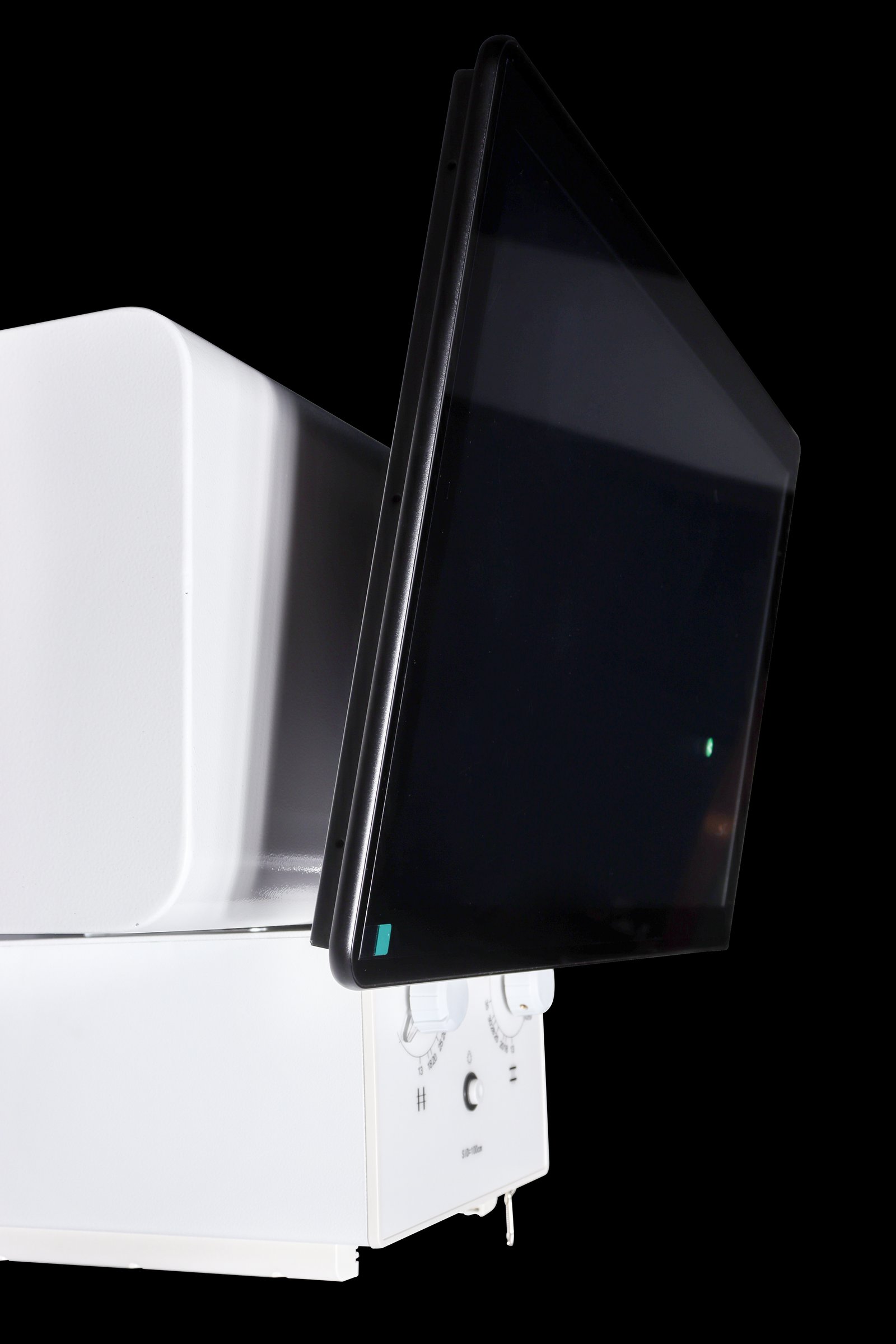
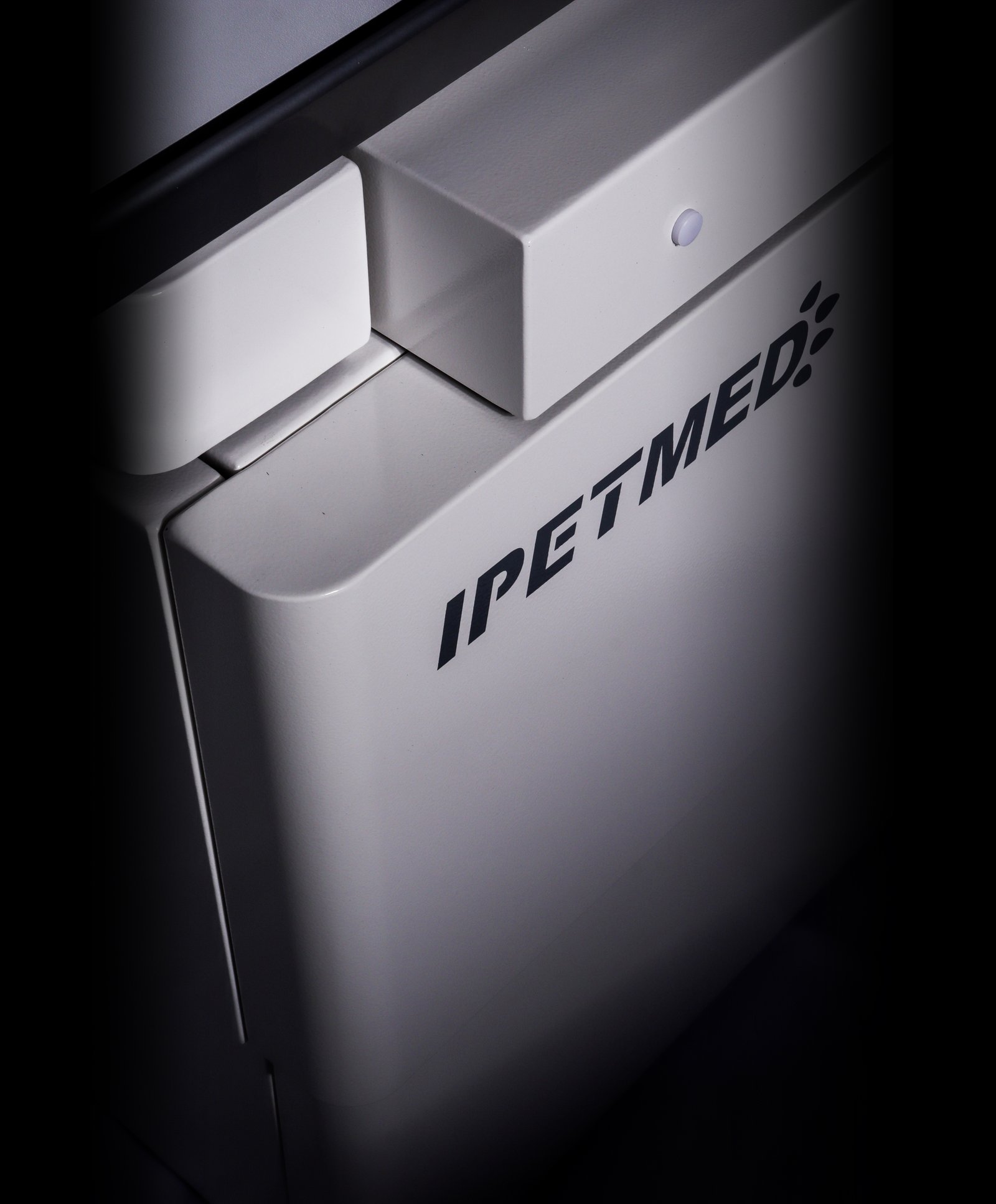
Overall dimensions and detail views: tube column, tube head and high-voltage assembly, four-way floating table, LED collimator, stainless-steel table edging, and touchscreen exposure controls.
System Specifications
| High-voltage generator | 32 kW high-frequency (veterinary), single phase 110/220 VAC |
| kV range | 40 – 150 kV |
| mA range | 10 – 400 mA |
| Exposure time | 1 – 10000 ms |
| X-ray tube | Canon E7239X |
| Tube max voltage | 125 kV |
| Focal spot | 1.0 / 2.0 mm (dual) |
| Anode input power | 21 kW (small focus) / 42.5 kW (large focus) |
| Anode heat capacity | 140 kHU |
| Patient table | 1400 × 700 mm, phenolic resin top, four-way float (X 75 ±10 mm / Y 200 ±10 mm) |
| Collimator | LED light source, 1.0 mm Al filtration at 75 kV |
| Workstation | Industrial PC, 23.8″ diagnostic monitor + 21.5″ tableside touchscreen |
| Power supply | Single-phase mains, no three-phase supply required |
Flat-Panel Detector (iLume 4343)
| Detector type / scintillator | a-Si / CsI |
| Active area | 43 × 43 cm (17 × 17″) |
| Pixel matrix | 3072 × 3072 |
| Pixel pitch | 139 µm |
| Spatial resolution | 3.6 LP/mm |
| A/D conversion | 16 bit |
| Min. detectable dose (RQA5) | 14 nGy |
| Image acquisition | 1 s |
| Data interface | GigE |
| Sealing | IP65 |
Software Configuration
| Acquisition workstation | In-house software with Caravaggio tone engine, body-size automatic exposure, collimator auto-detection, VHS cardiac measurement |
| AI imaging assistant | Draft report generation; veterinarian reviews and signs off (assist tool, not a diagnosis) |
| DICOM | DICOM 3.0 — Store, Print, and Worklist integration with practice-management software |
| Cloud imaging | Cloud image storage and cross-device access supported |
Clinical Applications
General canine and feline practice: orthopaedic, thoracic and abdominal radiography, pre-anaesthetic evaluation, and post-operative rechecks; routine imaging of small and medium exotic species.
For single-exposure full-spine or bilateral-hip studies in giant-breed canines, a 17 × 24″ large-panel variant is available on request. For dynamic studies (barium swallow, airway evaluation), see the iSmart-32Pro dynamic DR system.
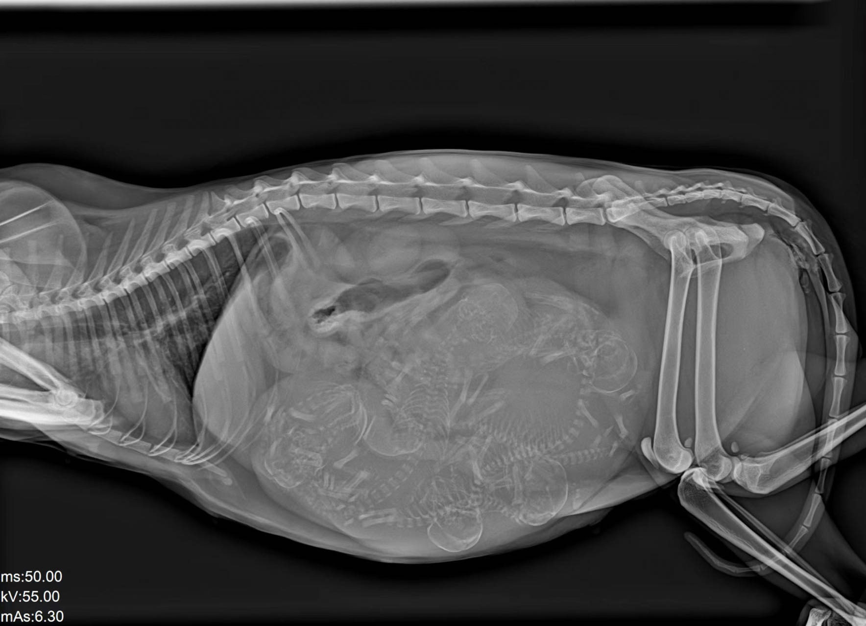
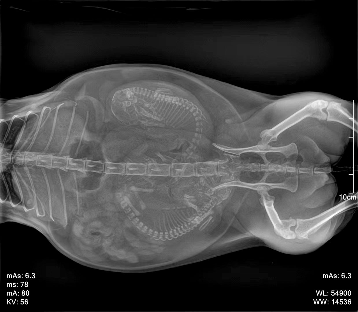
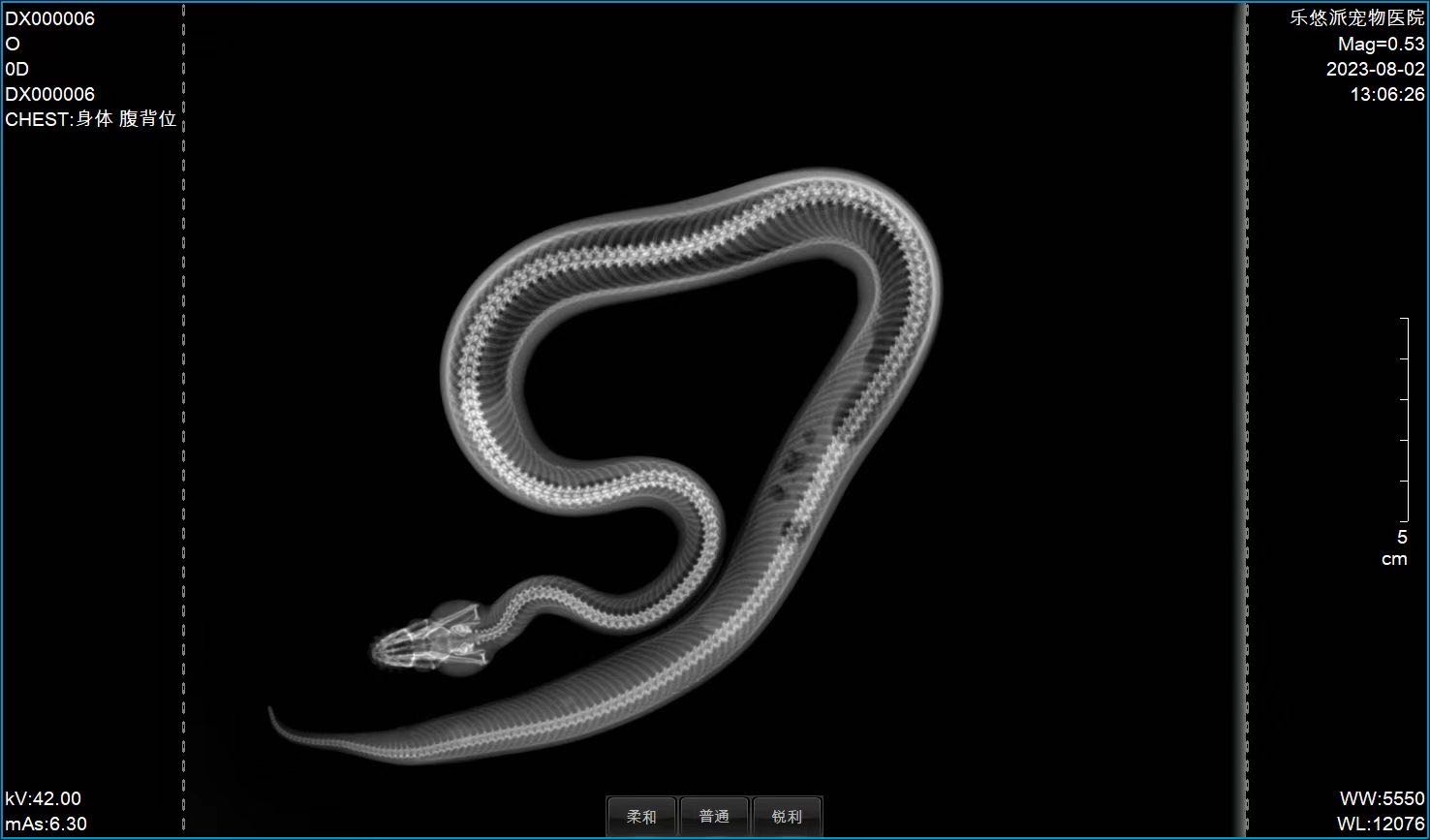
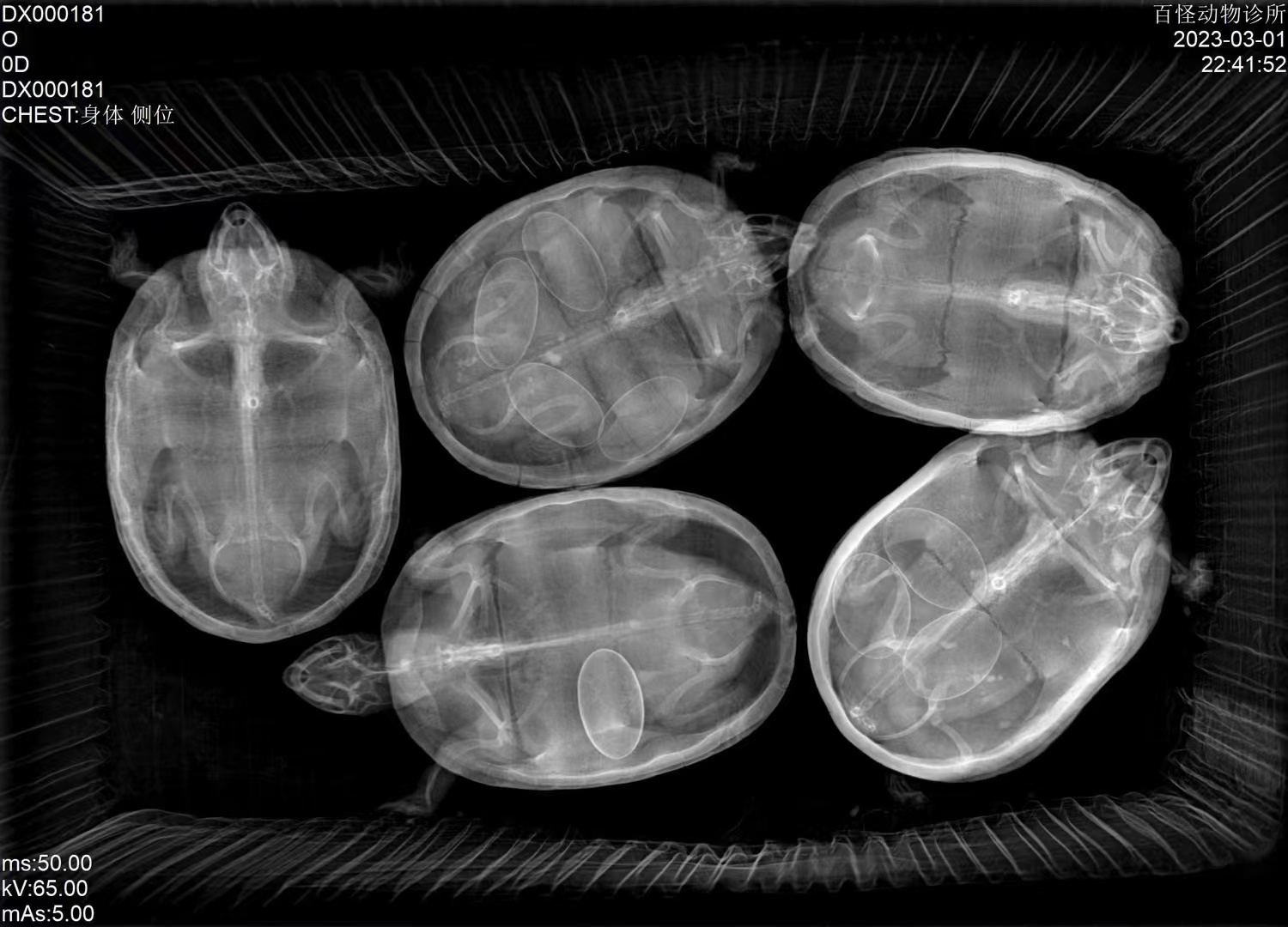
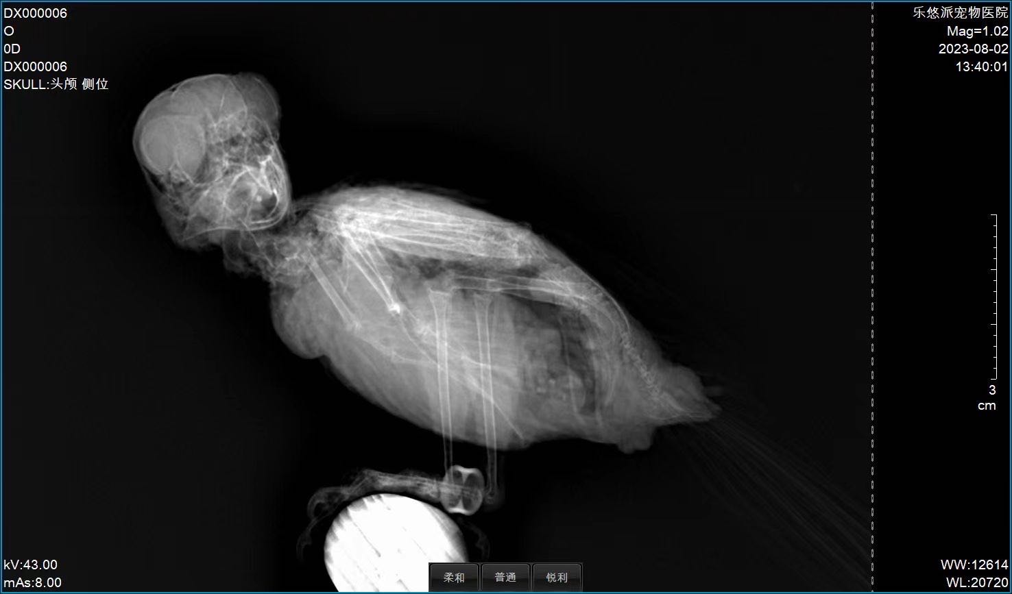
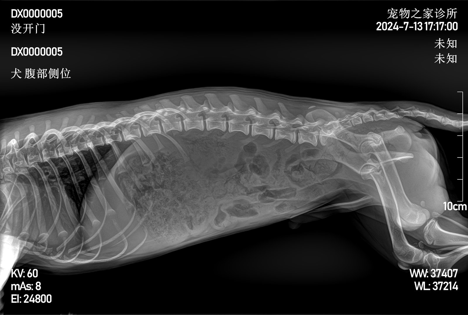
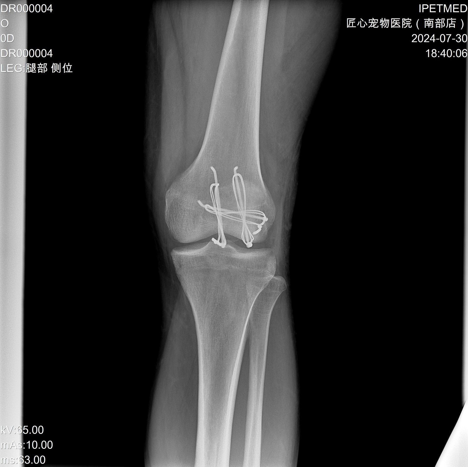
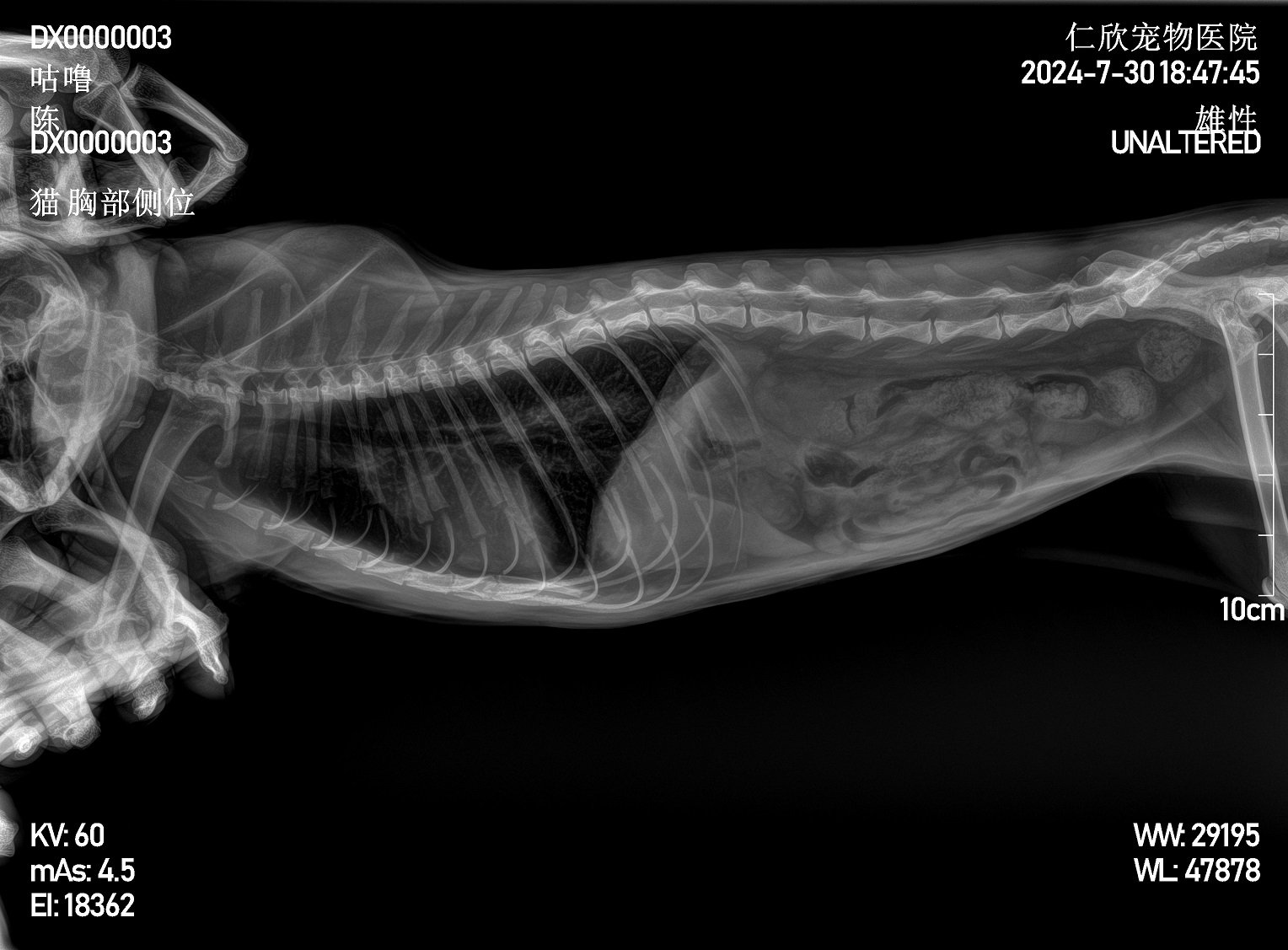
Clinical Imaging Samples
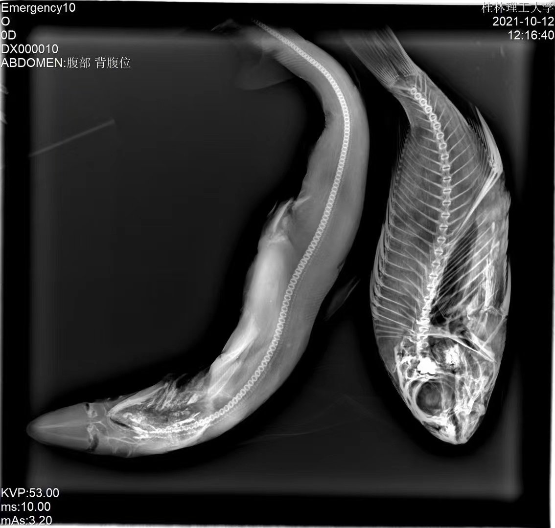
Representative radiographs from the iPet DR imaging chain across companion-animal and exotic species, illustrating image quality across body sizes and tissue contrast.
Workflow & Installation
Single-phase 110/220 VAC power, no dedicated three-phase supply required. Installation footprint accommodates the 1.4 m patient table with overhead tube column. Operating environment: 5–35 °C, 10–75 % RH. Engineer-led installation and on-site operator training are included.
iPet-400 platform installation and operator training walkthrough (English narration, 6 min)
Package Contents
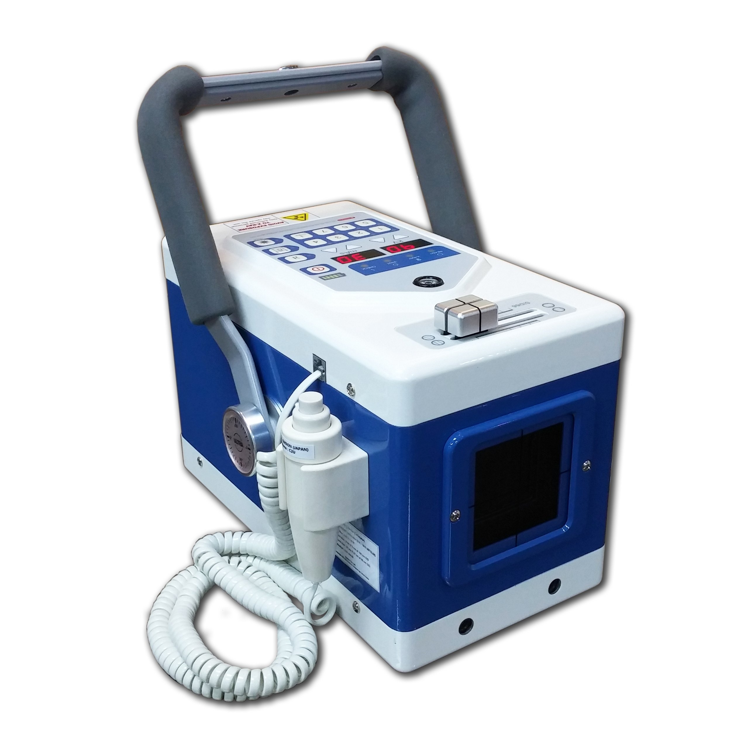
- 32 kW high-frequency generator + control box
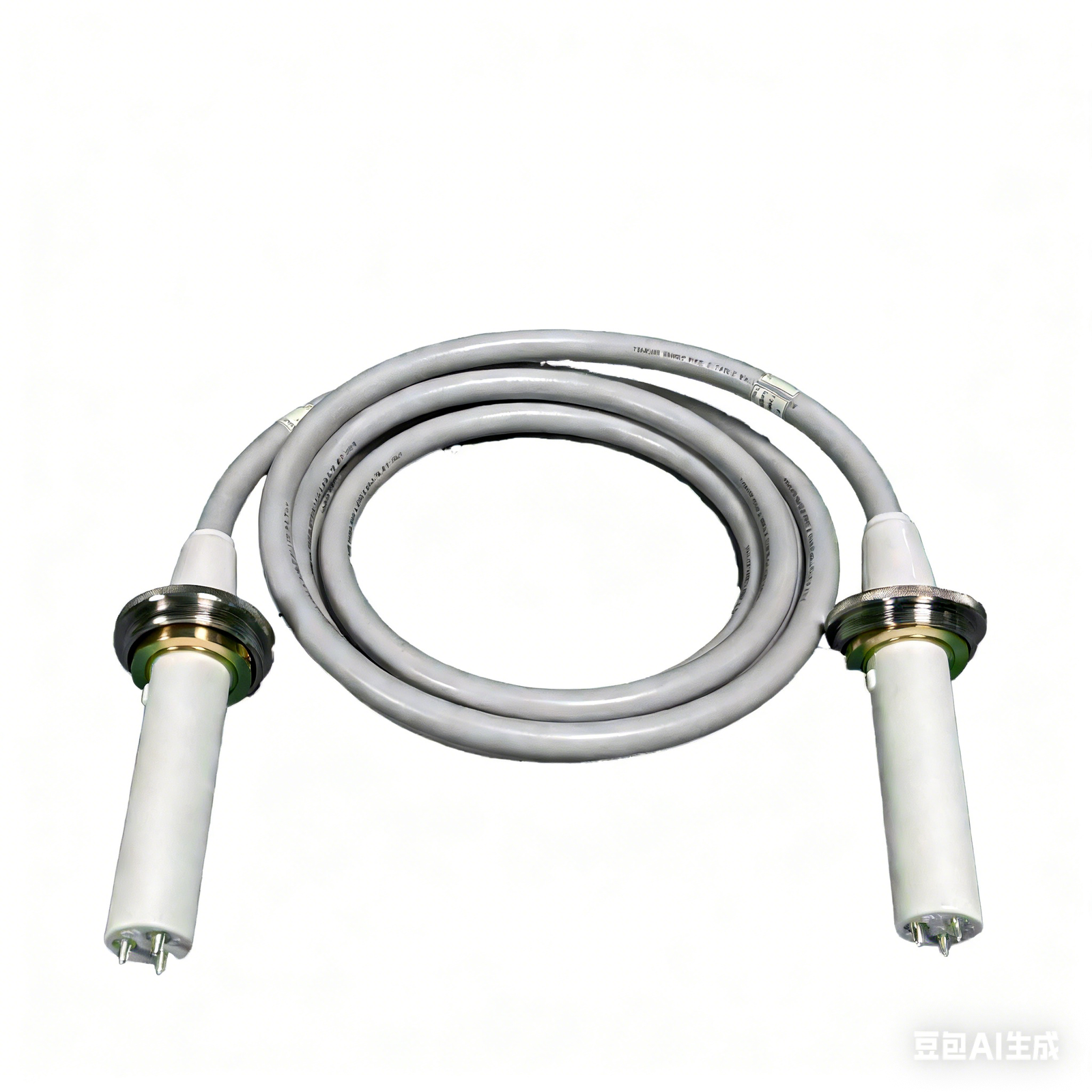
- X-ray tube assembly (Canon E7239X) + high-voltage cables
- iLume 4343 flat-panel detector (43 × 43 cm) + power / data adapter
- LED collimator
- Patient table (1400 × 700 mm) with four-way floating top
- Acquisition workstation: 23.8″ display, 21.5″ touchscreen, PC, software dongle
- V-shape positioning set (1 large + 1 small)
- Lead protection set: gown, hat, scarf, glasses, gloves
- Foot switch, remote control, network cable (10 m)
Downloads
Why Choose iPet-400AI
- Workflow efficiency — body-size automatic exposure keeps retakes low; AI report drafts cut routine reading time.
- Clinical accuracy — 32 kW high-frequency generator and Canon tube hold output steady through a twenty-exposure day.
- Practice economics — one manufacturer accountable for machine and software; cloud storage keeps images available across workstations and branch clinics.
Warranty & Support
Two-year warranty covering hardware and software. Extended-warranty service is available. Engineer-led on-site installation and operator training included.
FAQ
Q: Can the AI imaging assistant issue reports on its own?
No. The AI assistant generates a draft report only; the veterinarian reviews, edits, and signs off. It is an assist tool, not a diagnostic device.
Q: What is the iLume 4343 panel?
iLume is IWA’s own-brand detector line, manufactured by a specialist flat-panel factory and tuned at SDK level with the in-house acquisition software for a matched imaging chain.
Q: Can a giant-breed full spine be captured in one exposure?
The 43 × 43 cm panel covers routine views. For single-exposure full-spine or bilateral-hip studies, a 17 × 24″ large-panel variant is available on request.
Q: Does the system require three-phase power?
No. Standard single-phase mains is sufficient.
Q: What PACS / DICOM standards are supported?
The workstation is DICOM 3.0 compliant for Store, Print, and Worklist with most third-party PACS.

Reviews
There are no reviews yet.