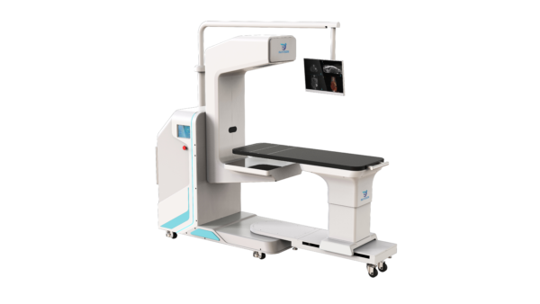
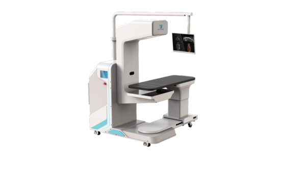
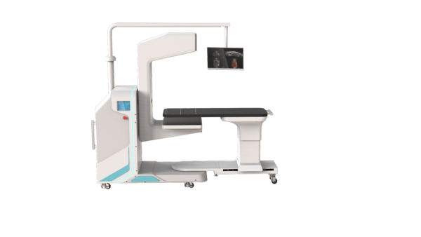
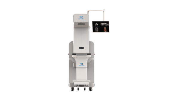
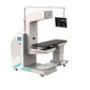
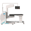
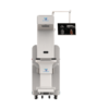
Cone-beam CT built around the operating table — a C-arm gantry with DSA and 0.16 mm slices for intraoperative imaging.
Product Overview
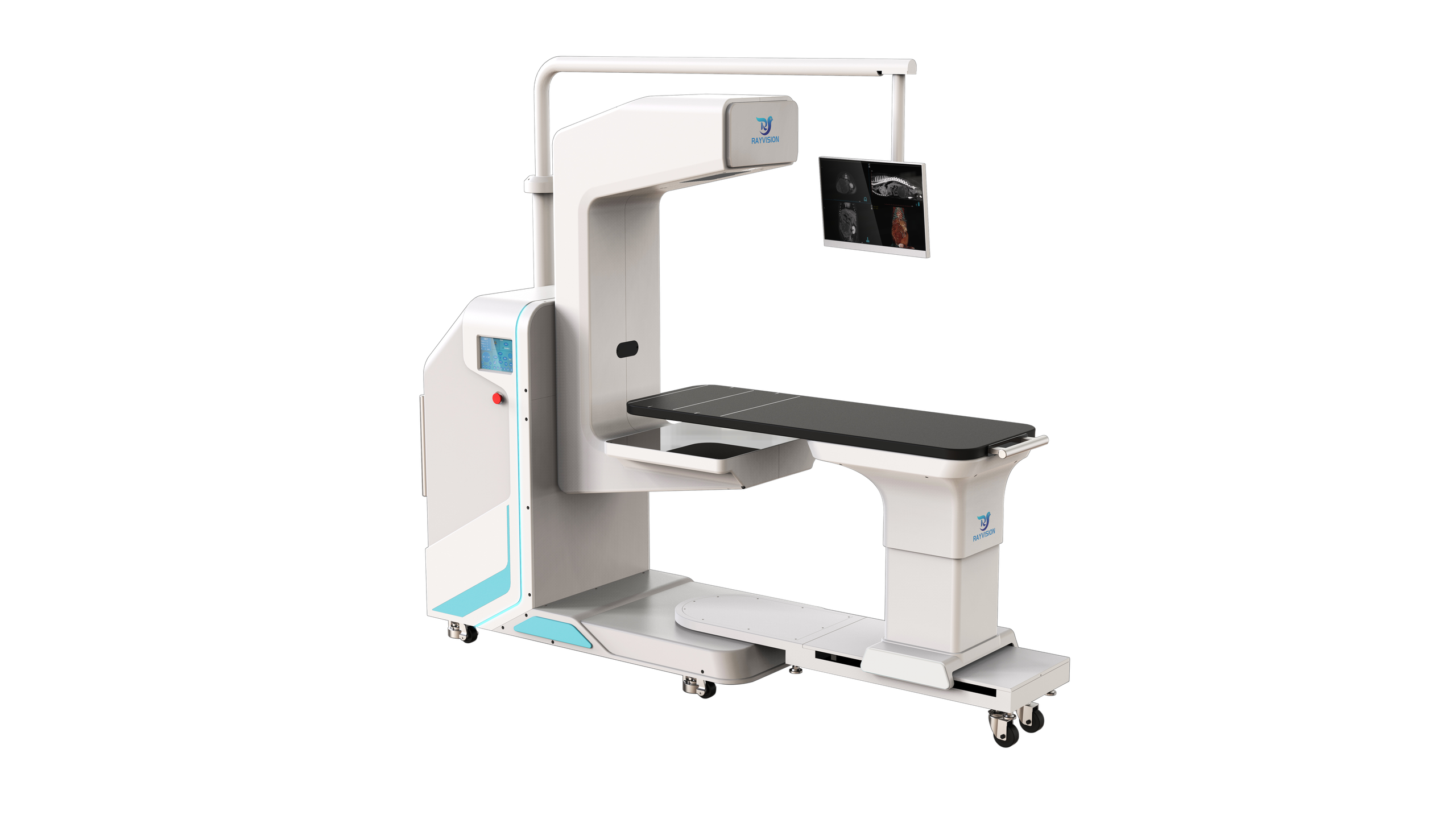
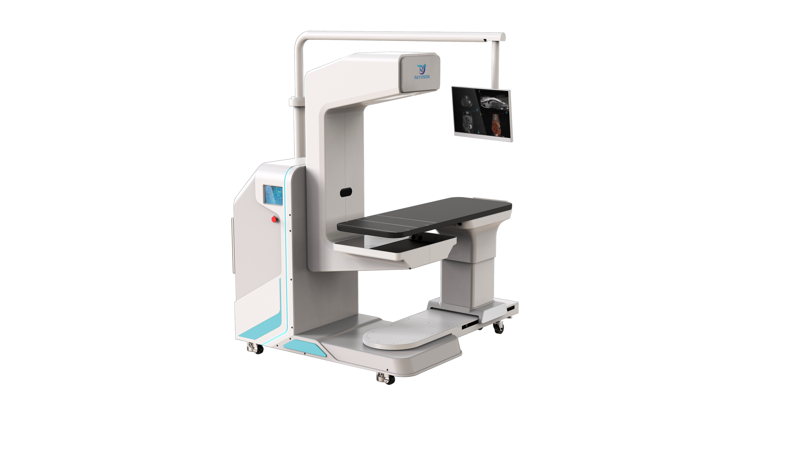
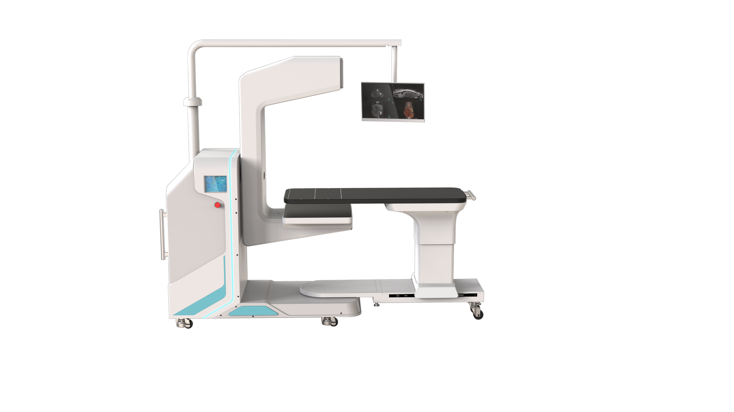
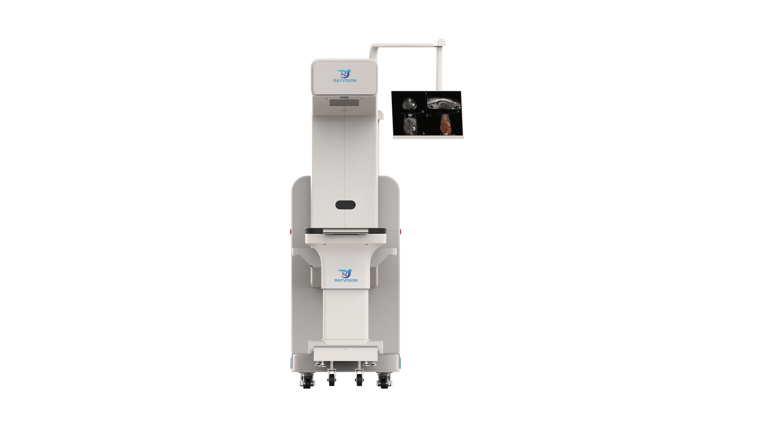
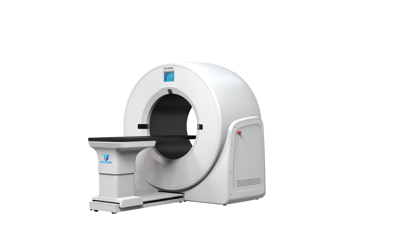
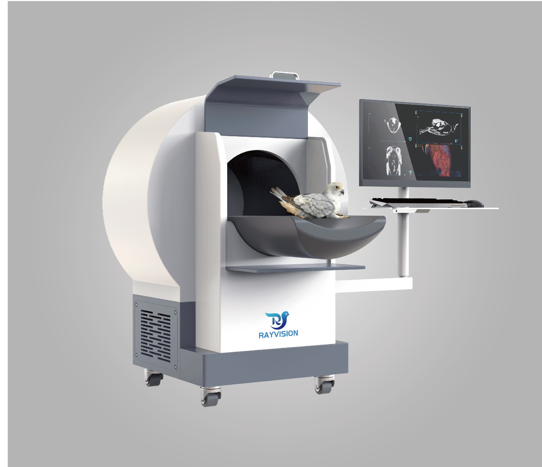
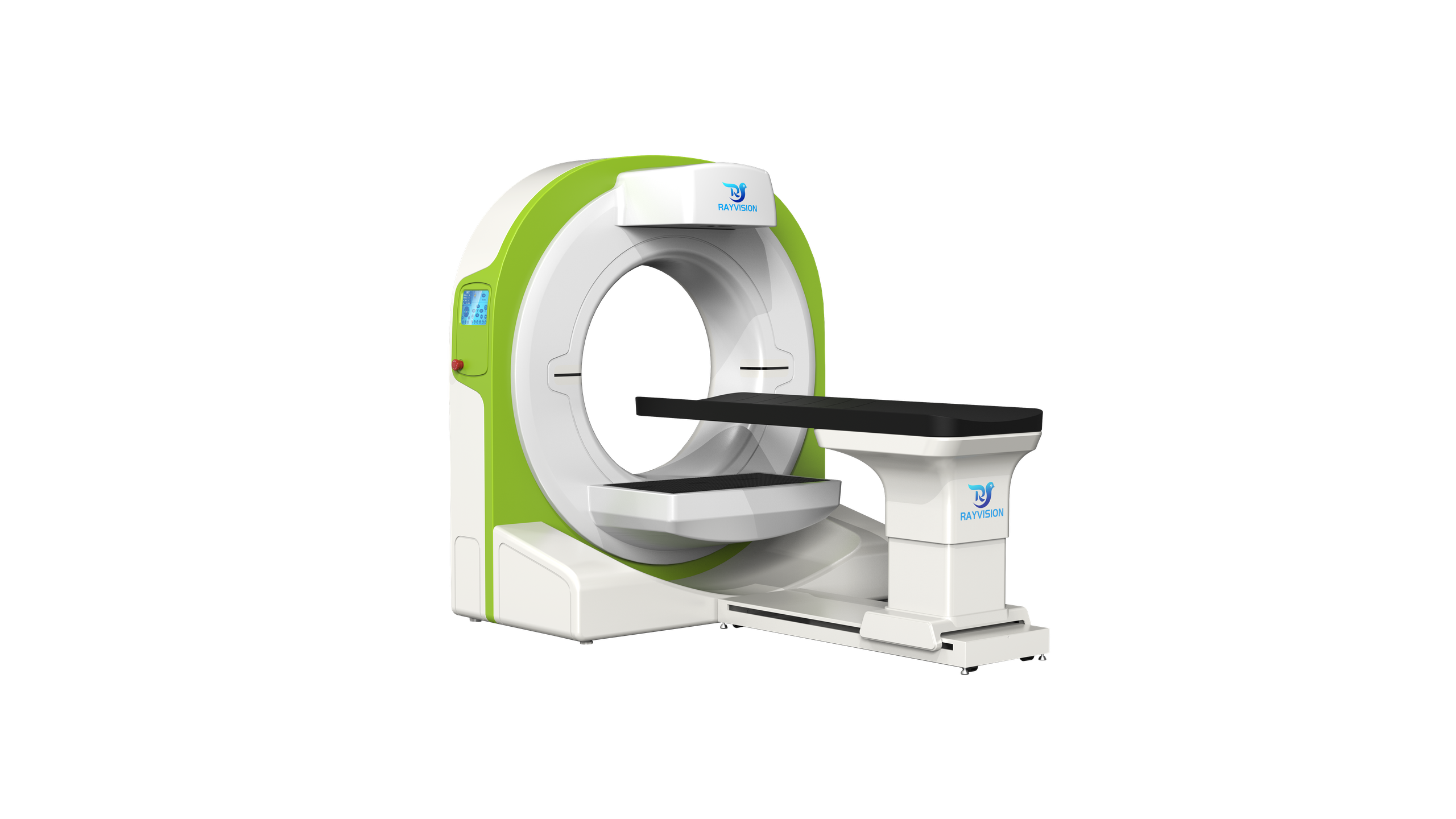
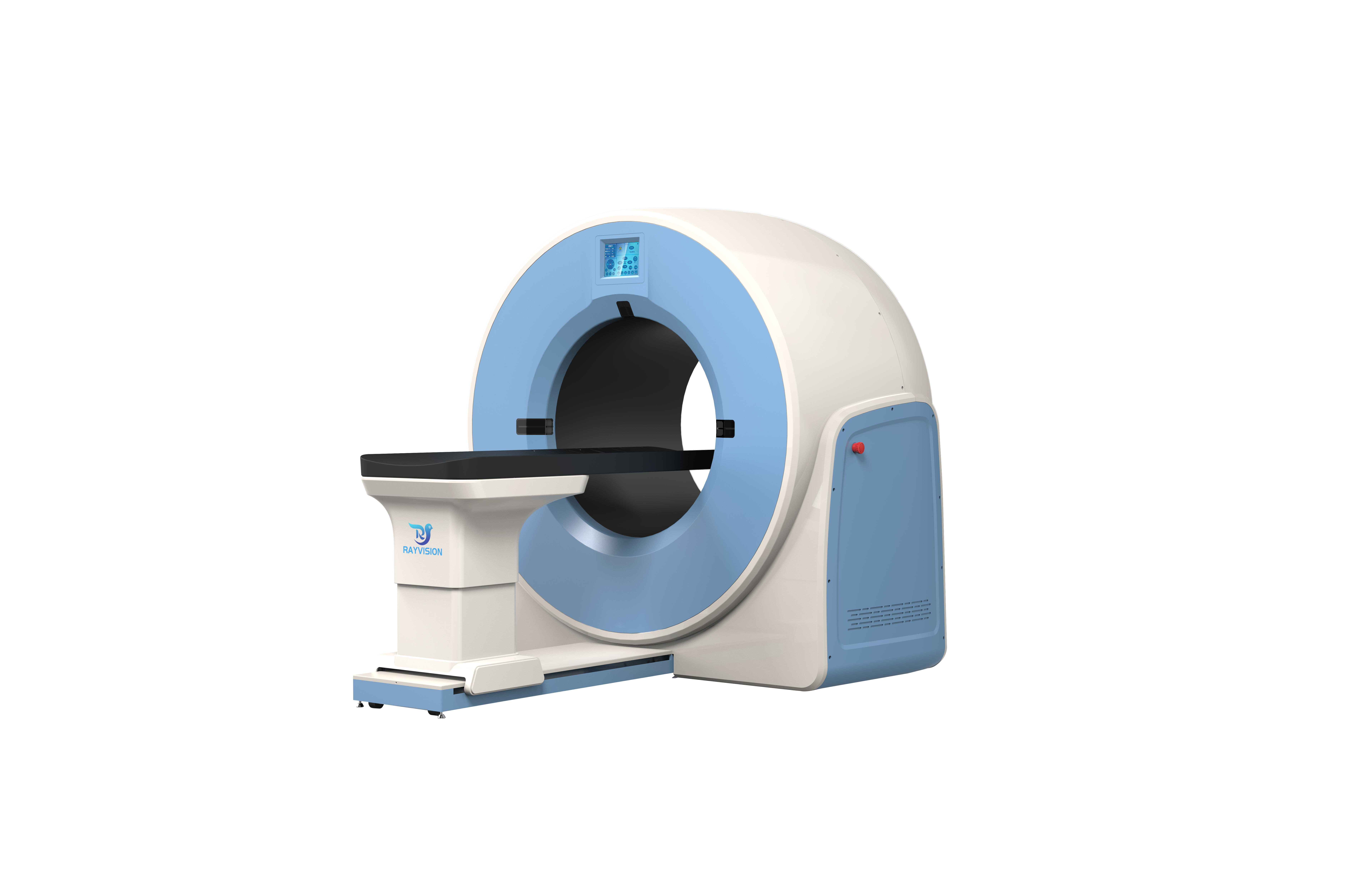
Vetopia 50 (Advance Series) is a C-arm veterinary cone-beam CT system from Rayvision Medical, designed for intraoperative imaging. The C-arm rotates freely through 360°, the patient table rotates to multiple angles, and an intraoperative monitor displays anatomy and images at the table side throughout the procedure.
The system combines CT, static DR, fluoroscopy and dental panoramic imaging, with DSA for interventional guidance. The CT field of view is 28 × 28 cm per scan; Vetopia 50 does not offer stitched extended-length scanning — for longer coverage, see Vetopia 70 (55 × 28 cm).
Key Features
- Flexible C-arm — 360° free rotation for additional viewing angles during surgery.
- Multi-angle table and intraoperative monitor — table rotation adapts positioning; the monitor shows anatomy and images in real time.
- 4-in-1 imaging — CT, static DR, fluoroscopy and dental panoramic.
- DSA — digital subtraction angiography shows vascular structures to guide interventional procedures.
- Intraoperative dynamic imaging — real-time capture of lesion changes during surgery.
- CT-AIR breath-hold platform — holds the patient at end-inspiration to eliminate motion artefact.
- Smart MAR — suppresses metal artefact around orthopaedic implants for post-operative review.
- 0.16 mm slices, 600 reconstructed slices — 0.14 mm minimum pixel size, 4.5 s scan.
Product Gallery
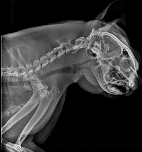
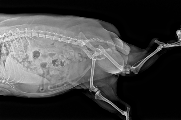
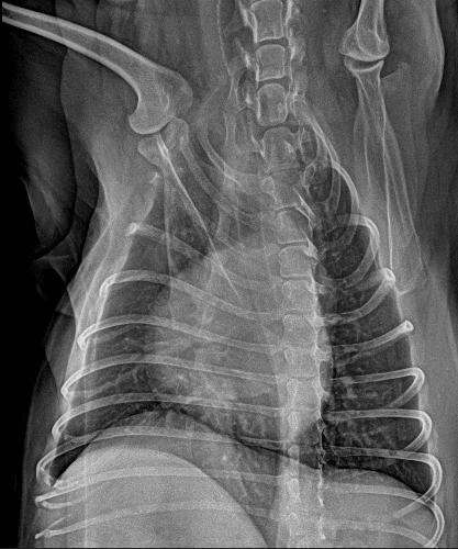
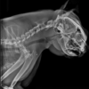
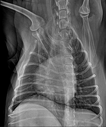
Clinical Imaging Samples
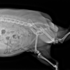
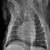
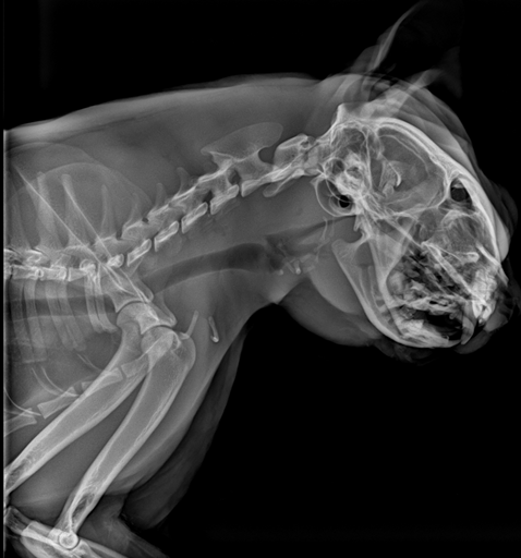
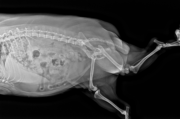
Reference radiographs acquired in DR mode.
Video
Vetopia 50 product introduction
Technical Specifications — Gantry
| Gantry structure | C-arm |
| Nominal power | 15 kW |
| Input power | (220 ±22) VAC |
| Tube heat capacity | 150 kJ |
| Maximum tube voltage | 120 kV |
| Tube power | 15 kW |
| Overall dimensions (drawing) | 2379 mm length × 1627 mm height (front view); 546 mm width (side view) |
Technical Specifications — Scan Parameters
| Reconstructed slices | 600 |
| Minimum slice thickness | 0.16 mm |
| Minimum pixel size | 0.14 mm |
| Detector matrix | 3072 × 3072 |
| CT scan time | 4.5 s |
| Maximum FOV (L × W) | 28 × 28 cm (11.02″ × 11.02″) |
| Stitched extended-length scanning | Not supported |
Technical Specifications — Imaging Modes
| CT | 0.16 mm slices, CT-AIR breath-hold, experience-triggered triple-phase contrast |
| DR | Edge enhancement, multiple processing algorithms, TPLO / VHS / TTA measurement tools |
| Fluoroscopy (dynamic DR) | Real-time dynamic enhancement, up to 1 minute continuous exposure |
| Dental panoramic | Full upper and lower arches and jaw from one scan |
| DSA | Digital subtraction angiography for interventional guidance |
Clinical Applications
| Region | Conditions |
|---|---|
| Head & neck | Cerebral oedema, brain haemorrhage, sinusitis, otitis, mastoid effusion, nasopharyngeal stenosis, tracheal narrowing |
| Dental | Periodontitis, periapical abscess, alveolar bone atrophy, oral cysts, tooth root resorption |
| Thoracic | Pulmonary oedema, pulmonary nodules, interstitial pneumonia, pulmonary inflammation |
| Abdominal | Abdominal wall inflammation, peritoneal effusion, urography, renal imaging, cholelithiasis, pancreatitis, renal sclerosis |
| Vascular | Abdominal triple-phase angiography, peripheral arterial thrombosis, vascular stenosis |
| Spinal | Myelography, disc protrusion, vertebral imaging, spondylosis, thoracic vertebral fracture |
| Oncology | Thyroid, nasal, ovarian, renal and adrenal tumours |
| Other | Arthritis, thoracic duct imaging, peritoneal calcification |
Clinical Case Highlights
- Renal infarction — Plain scan of the right kidney was unremarkable; after contrast, a cortical enhancement defect indicated partial right renal infarction in a patient presenting with anorexia, vomiting, dehydration and abnormal urination.
- Gallbladder mucocele — Homogeneous hepatic enhancement in the delayed phase; the gallbladder was filled with irregular soft-tissue-attenuating material with uneven wall thickening.
- Post-processing — Cardiac 3D rendering, vascular MIP and curved reconstruction of the thoracic duct.
Site Planning and Installation
Vetopia 50 is a fixed installation in a shielded room. The manufacturer’s layout drawing uses a 3000 × 3000 mm room with an 850 mm door.
Power and Environment
| Supply voltage | Single-phase (220 ±22) VAC |
| Supply internal resistance | ≤ 0.5 Ω |
| Frequency | 50 Hz / 60 Hz |
| Power capacity | ≥ 15 kVA |
| Power cable | 6 mm² |
| Shielded room | 3 mm lead equivalent |
| Operating temperature | 10 °C – 35 °C |
| Relative humidity | 30 % – 75 % RH, non-condensing |
| Atmospheric pressure | 700 – 1060 hPa |
| Wall clearance | ≥ 30 cm around the system |
Shielded Room Preparation
Room requirements from the manufacturer’s CT installation-conditions guide; a site survey confirms the final layout.
| Room area | Minimum 3 × 3 m; 3.5 × 3.5 m recommended |
| Room height | ≥ 2 m |
| Door opening | ≥ 1 m wide for whole-unit entry (≥ 0.85 m with the main-unit covers removed); ≥ 1.8 m high |
| Wall shielding | 240 mm clay brick + 20 mm protective coating, or any construction equal to 3 mm lead; concrete walls ≥ 240 mm |
| Floor / ceiling shielding | Ground floor: ceiling 3 mm Pb. Intermediate floors: floor and ceiling 3 mm Pb. Top floor: floor 3 mm Pb |
| Floor | Level concrete, foundation depth > 120 mm, flatness ≤ 2 mm, load capacity > 500 kg/m²; ground floor preferred |
| Delivery route | Ramps < 5°; use a forklift or lifting equipment for steeper access |
| Observation window | Standard lead glass 400 × 600 × 15 mm in a 405 × 605 mm opening, supported by 40 × 60 mm square tube |
| Cable entry | Ø 50 mm hole near the operator console |
| Electrical | Dedicated circuit breaker; voltage stabiliser if mains deviates more than ±10 %; UPS where outages are frequent |
| Signage | Radiation warning sign and exposure indicator light outside the room door |
Workflow Notes
- The C-arm and multi-angle table are positioned around the surgical field; the intraoperative monitor keeps images at the table side.
- The standardised viewing and reporting system generates templated reports for routine examinations.
- Each CT acquisition covers a 28 × 28 cm field of view; plan full-body studies accordingly.
Package Contents
- 1 × C-arm gantry
- 1 × Multi-angle patient table
- 1 × Intraoperative monitor
- 1 × Acquisition and reconstruction workstation
Downloads
Product brochures and technical documentation are available on request — contact IWA.
Why Choose Vetopia 50
- Workflow efficiency — Imaging comes to the operating table: C-arm rotation, table rotation and a table-side monitor avoid moving the patient mid-procedure.
- Clinical accuracy — DSA and intraoperative dynamic imaging show vascular anatomy and lesion change in real time; Smart MAR clears implant artefact.
- Practice economics — One C-arm system covers intraoperative CT, fluoroscopy, DR and dental panoramic imaging.
Warranty and Support
1-year whole-system warranty. Extended warranty is available for purchase — contact IWA for terms and service coverage in your region.
Installation is carried out against the manufacturer’s installation manual: the main unit and patient table are connected, the console and workstation are cabled, and scanning, reconstruction, fluoroscopy and DR acquisition are verified before handover.
FAQ
Q: How does Vetopia 50 differ from Vetopia 70?
Vetopia 50 has a 28 × 28 cm CT field of view, 600 reconstructed slices and no stitched scanning. Vetopia 70 extends the field of view to 55 × 28 cm with 800 slices, and adds time-triggered triple-phase capture and database-assisted reporting.
Q: Is it suitable for interventional procedures?
Yes. The C-arm design, DSA and intraoperative dynamic imaging are intended for surgical and interventional guidance.
Q: What room does it need?
A shielded room of 3 mm lead equivalent (manufacturer layout: 3000 × 3000 mm), a single-phase (220 ±22) VAC supply of ≥ 15 kVA on a dedicated breaker, and 30 cm clearance around the system.
Q: Does it include dental imaging?
Yes. Dental panoramic mode captures the complete upper and lower arches and the jaw from one scan.
Q: What warranty applies?
1-year whole-system warranty, with extended warranty available for purchase.
Reviews
There are no reviews yet.