The 15% Rule: When Thickness Changes, How Your Settings Follow
What do kV and mAs each control? How do you adjust when the chart has no row for this thickness? The most famous rule of thumb in radiographic technique, explained once.
 Veterinary imaging · Core software built in-house
The 15% Rule:
When Thickness Changes, How Your Settings Follow
What do kV and mAs each control? How do you adjust when the chart has no row for this thickness? The most famous rule of thumb in radiographic technique, explained once.
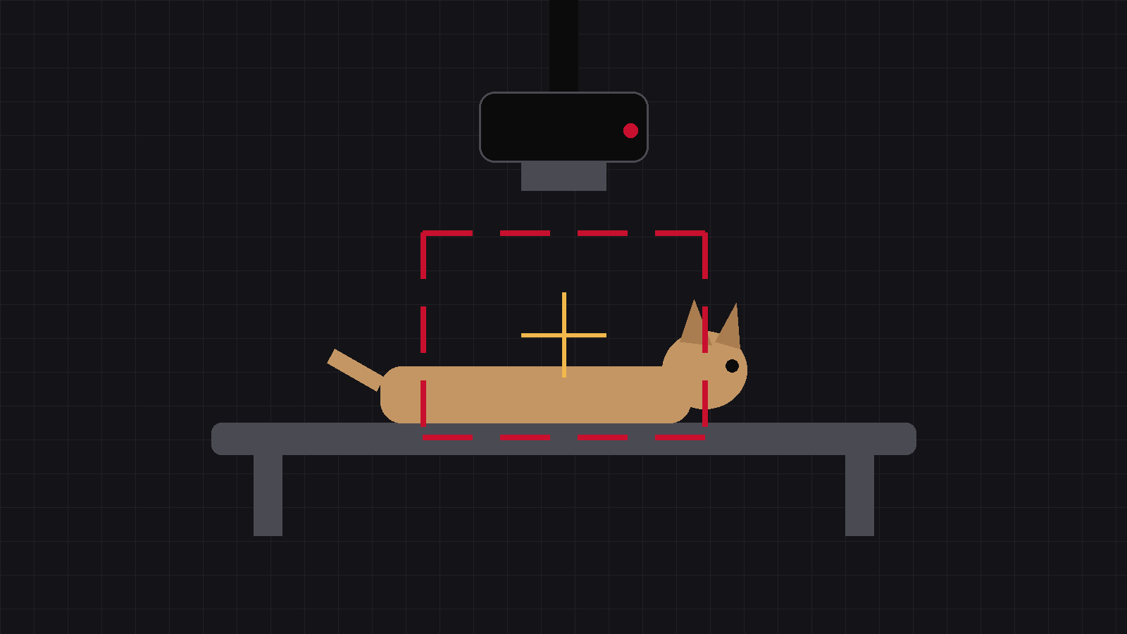
Last issue we said: measure the thickness, then look it up in the chart. But the chart cannot cover everything — the dog is two centimetres thicker and there is no such row; the image came out slightly pale and needs a touch more; or you want stronger contrast between bone and soft tissue. Which knob do you turn, and by how much? The answer lies in the division of labour between two knobs.
01First, know your two knobs
This is the foundation of everything:
kV (tube voltage) controls penetration
The higher the kV, the “harder” the beam — the better it gets through thick body parts. At the same time, high kV softens the image contrast: a longer grey scale with more shades, but less difference between bone and soft tissue.
mAs (tube current × time) controls quantity
The higher the mAs, the more X-rays you send — the image gets “enough”. It changes neither penetration nor contrast, only how much signal arrives: that means the amount of noise, and the dose the animal receives.
One line to hold on to: kV decides whether the beam gets through and how strong the contrast is; mAs decides whether there is enough signal and how noisy the image is.
02The 15% rule: converting between the knobs
Now for the most famous rule of thumb:
Raise kV by about 15% and, as far as the detector is concerned, the effect roughly equals doubling the mAs. Conversely, dropping kV by 15% is roughly the same as halving the mAs.
What the rule buys you is a way to “convert” between the two knobs. Say an image taken at 60 kV needs more exposure: you can double the mAs, or raise the kV to about 69. Both routes give you an image that is “enough” — but the outcomes differ: the kV route softens contrast a little and may actually lower the dose; the mAs route keeps contrast unchanged and doubles the dose outright.
The clinical preference
Thick regions where you want soft-tissue detail (chest, abdomen) — lean towards raising kV. Bone work where you want crisp contrast — adjust mAs and leave kV alone.
And when body thickness clearly changes? Move kV first to bring penetration back in line, then fine-tune with mAs — the exact step sizes come from your machine’s technique chart and your own records. As we said last issue: your own clinic’s chart is the real answer.
03Three working disciplines
Change one knob at a time
Move kV and mAs together, and when something changes you will never know which knob deserves the credit — or the blame.
Discipline 2: kV follows thickness first, mAs fine-tunes after
A change in thickness is a penetration problem. Solve penetration first, then worry about quantity.
Discipline 3: write it down
When a change produces a good image, write those settings into your chart so next time you just look them up.
Decide what you lack before you reach for a knob, and your settings stop being guesswork.
kV +15% ≈ mAs ×2.
Change one knob at a time.
Source:The 15% rule is a general rule of thumb in radiographic technique (raising kVp by about 15% ≈ doubling receptor exposure); for actual settings, always follow your own machine’s technique chart.
IWA Medical · We build DR systems and write our own core software
Questions about veterinary DR imaging? Technical questions go straight to our engineers.