
Why DR Uses Less Dose Than Film Did
Everyone says DR uses less radiation than film — less where? The position first: this is a difference between generations, not between brands; clinics still on film or CR are using what was the best technology of its day. The saving sits in three places: the detector catches more (higher DQE), latitude rescues near misses (a slightly off technique no longer means a repeat), and the instant image ends the habit of buying insurance with dose. But the dividend does not collect itself — technique chart, exposure index, collimation, first-time-right.
 Veterinary imaging · Core software built in-house
Why DR Uses Less Dose
Than Film Did
For the same diagnostic image, a DR system needs less dose than the film era did. Less where, exactly? Three places: the detector catches more, latitude rescues near misses, and an instant image removes the need for a safety margin. One thing first, though — this is a difference between generations, not between brands.
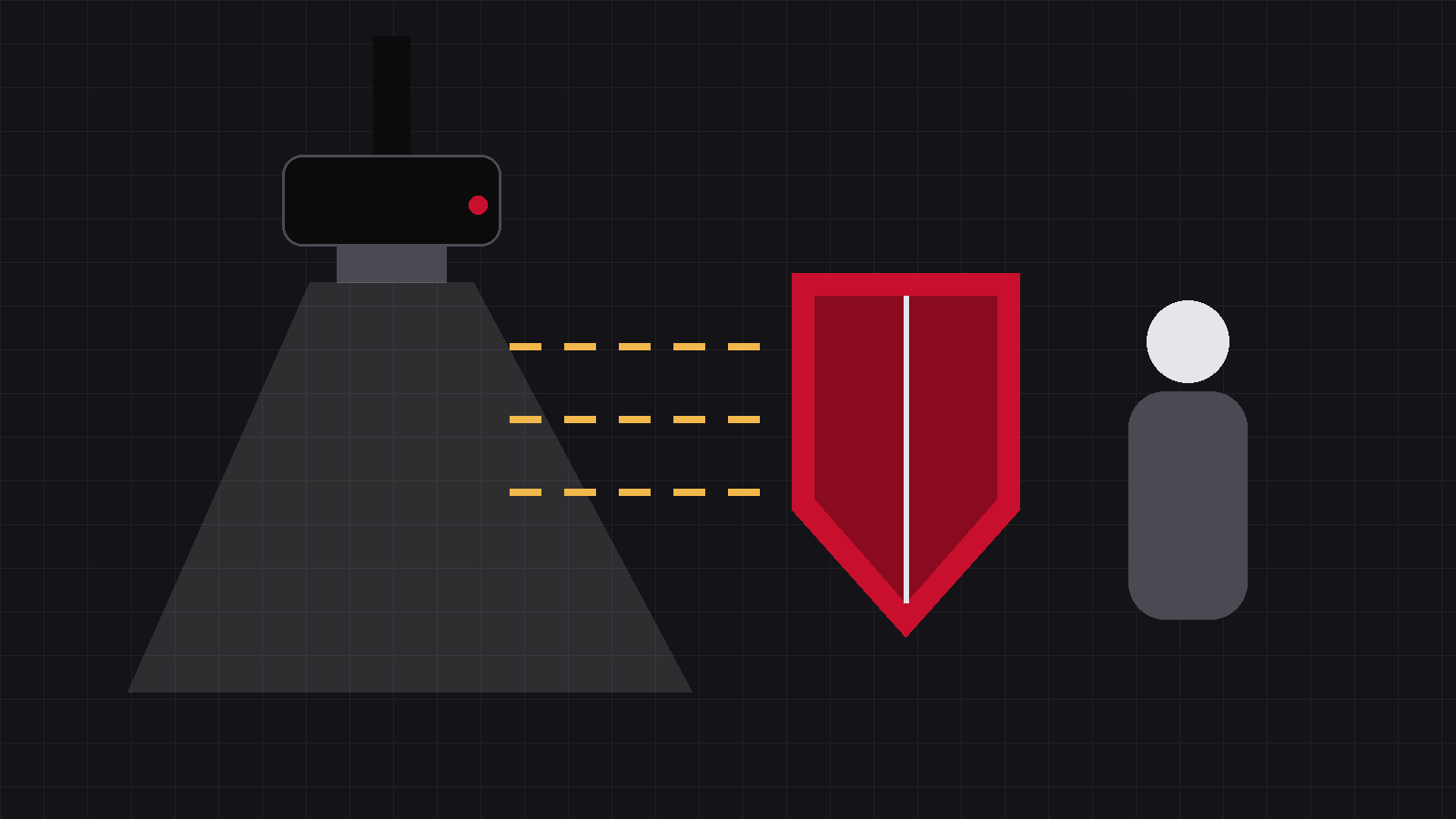
It gets asked often enough to deserve a careful answer: everyone says DR uses less radiation than film — less where? Answered badly, the question turns into a scare story. So the position goes first: this issue is about a difference between generations of technology, not between brands.
01Getting the position straight
Clinics still working with film or CR are using what was the best technology of its day. It found countless fractures, foreign bodies and cases of pulmonary oedema, and it saved lives. There is nothing in it to look down on — nobody mocks a ten-year-old phone for not having an edge-to-edge screen.
But the generational difference is real, and it does not live in some vague notion of sharpness. It lands on a very concrete number: dose.
Two generations of technology, not two brands
Everything below is true of any competent digital radiography system, whoever built it. Dressing up a generational advantage as a house advantage would be dishonest.
02Where the saving comes from: three of them
The lower dose is not down to one clever feature. Three things stack up.
① The detector catches more: less radiation passes through for nothing
The measure of how well a receptor catches what reaches it is DQE (detective quantum efficiency): of the information the beam carries out of the patient, how much the detector actually turns into image.
In the film-screen generation that figure was low — a great deal of radiation passed straight through to no purpose, contributing nothing to the image while still being delivered to the animal. Digital flat panels, caesium iodide (CsI) panels in particular, have a substantially higher DQE.
Catch more, and you need to send less. The same diagnostic-quality image is obtained at a lower dose. That is what the abstract-sounding line — detector efficiency is itself a form of protection — means in practice.
② Latitude rescues near misses: a slightly off technique no longer means a repeat
Film has a narrow exposure window: one step too high and the image is black, one step too low and it is blank, with a thin usable gap in between. A DR system’s dynamic range is far wider, and a small error in technique can be pulled back into a readable image in processing.
All of the dose saved by this one sits in retakes. Issue 11 went through the real bill for a retake, and the conclusion was blunt: every retake avoided is dose genuinely saved — saved twice over, in fact, since a retake means the same region takes a full exposure all over again.
③ The image appears at once: no more insurance
This is the easiest one to overlook, and it is the one that changes human habits.
In the film era, adding a little was the rational choice. You only learned whether the image had worked once the darkroom had finished with it, and by the time you saw it was underexposed the animal had gone home — someone had to phone the owner and ask them to bring it back in. To avoid that scene, an experienced radiographer’s hand quietly went up a step. Dose was being spent on insurance.
DR removes the reason for it. Press the switch, and two or three seconds later the image is on the screen; you know there and then. With no insurance to buy, technique can be set to what the image actually needs.
Put the three together and it comes to one sentence: less radiation has to be sent, less of what is sent goes to waste, and fewer attempts go wrong. That is the generational dividend.
03But the dividend does not collect itself
Here comes the cold water.
Wide latitude is a good thing, and it has a side effect: technique can drift well above what was needed and the image still looks fine. Nobody is punished, so nobody notices. Settings get a little more generous each time, dose climbs quietly, and the profession has a name for it: dose creep.
Equipment supplies the capability, not the result
DR makes low dose possible. Whether that possibility is realised still comes down to how the machine is operated. You need the technology and the technique. Neither one alone.
Four unglamorous habits are what turn the dividend into real dose reduction:
Work from the technique chart
Set exposures by measured body thickness, not by feel. That extra step added by feel is where dose creep begins.
Read the exposure index (EI)
EI is the system telling you how much that image actually received. A high EI means the technique was generous — the image will not show it, the EI will.
Collimate
Bring the field down to what you actually need to see, and irradiate nothing beyond it. This costs nothing and takes nothing away from the diagnosis.
Get it right first time
Positioning, restraint, and timing with respiration. Get those three right and the retake rate falls on its own. Every retake saved is a whole exposure’s worth of dose.
04When someone asks whether moving to DR is worth it
Say it without scaremongering and without exaggeration: it is not that film was harmful, it is that the new generation genuinely uses less. Less through detector efficiency, less through fewer retakes, less because nobody has to buy insurance with dose.
One aside: because the two generations work on different logic, the old rule of thumb that underexposure makes an image pale does not transfer to DR. That one has an article of its own, so it is left aside here.
The three savings come from the equipment; collecting them comes from people. Chart, EI, collimation, first-time-right — do those four and the generational dividend actually reaches the animal.
DR’s lower dose comes from three savings: the detector catches more, latitude rescues near misses, and an instant image ends the safety margin.
The dividend is collected by good technique, not automatically.
Source:DQE (detective quantum efficiency) and dynamic range are general concepts in imaging physics; for the definition of DQE see the IEC 62220 series of standards. Dose creep is an established term in radiographic practice. Differences in detector efficiency between generations are described here qualitatively only: no figures are quoted, no brand comparison data is used, and nothing here is a performance claim for any particular system.
IWA Medical · We build DR systems and write our own core software
Questions about veterinary DR imaging? Technical questions go straight to our engineers.