
Where CBCT Truly Shines: Seeing the Structures Buried in Overlap
We build DR systems, and this article pulls no punches: dental, skull and maxillofacial, complex orthopaedics, anything that needs 3D — where CBCT is genuinely strong, and its trade-offs in the same breath.
 Veterinary imaging · Core software built in-house
Where CBCT Truly Shines:
Seeing the Structures Buried in Overlap
We build DR systems, and this article pulls no punches: dental, skull and maxillofacial, complex orthopaedics, anything that needs 3D — where CBCT is genuinely strong, and its trade-offs in the same breath.
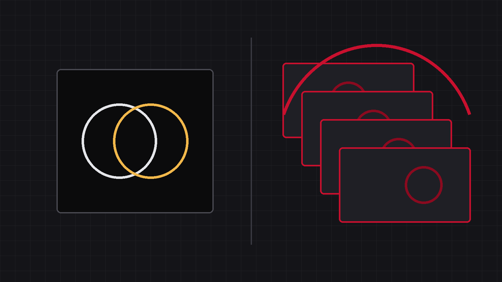
In Issue 1 we said: “DR sees a projection, CBCT sees slices.” This time we spell out what CBCT is genuinely good at — we build DR, and this article pulls no punches. Only when you know exactly where it is strong can you tell which cases to refer and which not to. Its whole skill comes down to one sentence: structures hidden behind other structures — it can peel them apart.
01The four areas where CBCT is genuinely strong
Roots, periodontal bone, unerupted teeth
Tooth roots, periodontal bone, alveolar bone loss, unerupted teeth, periapical lesions — on a plain radiograph these always overlap the neighbouring teeth and the jaw. Dog and cat molars are multi-rooted: on film, three roots collapse into one shadow. CBCT cuts through slice by slice, so each root is seen on its own. Exotics are even more telling: rabbit tooth roots grow deep into the jaw — film gives a rough idea, tomography shows exactly how far they have gone. If your clinic sees a lot of dental cases, CBCT’s value here is real.
The most overlapped region of the body
Nasal masses, the tympanic bullae of the middle ear, the temporomandibular joint, skull fractures — the skull is the most densely packed, most heavily overlapped region there is; on film, left and right, front and back are all pressed together. Slices pull them apart, and a 3D reconstruction lets you rotate the skull and look.
How many pieces, and where each one sits
Intra-articular fracture fragments, the small bone fragments of elbow dysplasia, vertebral malformation and stenosis, pre-surgical planning of complex pelvic fractures — film tells you “there is a fracture”; tomography tells you how many pieces, where each one sits, and from which direction to place the implant. Bone is a high-contrast structure, and CBCT’s spatial resolution on it is excellent — the detail is there.
Data a 2D film cannot provide
Surgical planning, 3D-printed guides, measuring angles and volumes — these need three-dimensional data by their very nature, and a two-dimensional projection cannot provide it.
These four areas are what DR cannot replace. When such a case comes in and the radiograph does not answer the question, refer it for tomographic imaging — do not push on regardless.
02The weak points, in the same breath — that’s only fair
It trades time, dose and investment for 3D without overlap
Soft-tissue contrast is not its strength — evaluating thoracic and abdominal organs remains the territory of DR and conventional CT; a scan takes tens of seconds and the animal must hold perfectly still, which usually means sedation or anaesthesia; the dose is higher than a single DR exposure; and the room, shielding, purchase and maintenance are all heavier than DR. Strength and weakness are two sides of the same coin.
03For the practice owner, the real question
Of the four kinds of cases above, how many do you see in a month? And how many of those were radiographed on DR, still left unanswered, and had to be referred out? That number is the starting point for the maths that comes later in this series — the dose maths, the money maths, the utilisation maths.
And to be plain about where we stand: we build DR — and when a case needs tomography, that is exactly what we tell our customers.
Keep those numbers — they are exactly what the “is a machine worth it” maths later in this series will run on.
Structures buried in overlap, bone in 3D detail —
that is where CBCT is simply strong. When it’s time to refer, refer.
Source:This article describes application scenarios and cites no external data; whether to refer any individual case for tomographic imaging rests with the attending veterinarian.
IWA Medical · We build DR systems and write our own core software
Questions about veterinary DR imaging? Technical questions go straight to our engineers.