
CBCT or DR? One Sees the Plane, One Sees the Slices
More and more practice owners are asking: “should I buy a CBCT?” Neither machine is “more advanced” — they simply see different things. One sees the plane, one sees the slices.
 Veterinary imaging · Core software built in-house
CBCT or DR?
One Sees the Plane, One Sees the Slices
More and more practice owners are asking: “should I buy a CBCT?” Before answering, get one thing straight: neither machine is “more advanced” — they simply see different things. One sees the plane, one sees the slices.
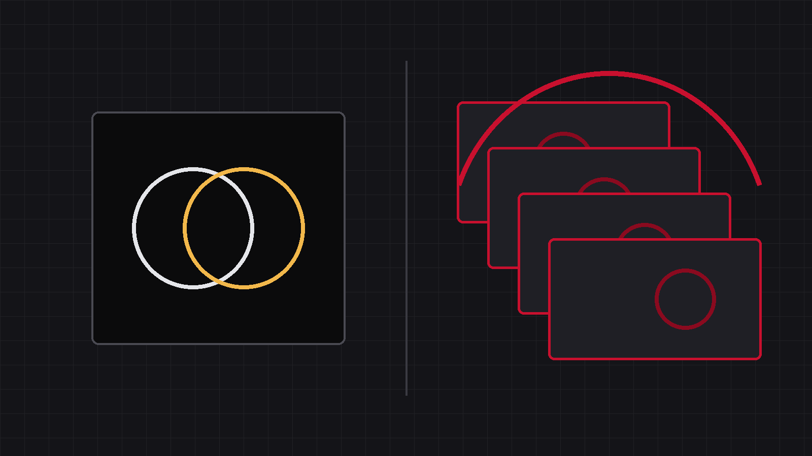
In the last couple of years, more and more practice owners have been asking: “Should I invest in a CBCT?” Before that question, there is another one to answer first: what exactly separates CBCT from DR? Not “which is more advanced” — they simply see different things.
01One flattens the thickness, one cuts the body open
The whole thickness, flattened onto one image
X-rays pass through the body from one side and land on a flat-panel detector on the other. The entire thickness of the body is compressed onto a single image — ribs, heart, lungs and spine all stacked front-to-back. The gain: one exposure, tens of milliseconds, one image showing the whole picture. The price: superimposition — two structures sitting one behind the other cannot be separated on a single plane.
One rotation, a 3D volume you can cut open
The tube and detector orbit the animal once, capturing hundreds of projections from different angles; a computer reconstructs them into a 3D volume you can slice layer by layer or rotate freely. The gain: no superimposition — hidden structures are peeled apart. The price: it takes tens of seconds, the animal must stay perfectly still (usually sedation or anaesthesia), the dose is higher than a single DR shot, and the machine and room require a much bigger investment.
02Each owns ground the other cannot take
So neither replaces the other — each has home ground the other cannot cover:
| What you need to see | Where DR shines | Where CBCT shines |
|---|---|---|
| Thorax & abdomen, soft-tissue overview | ✅ First choice — one image shows it all | Weak soft-tissue contrast; not its strength |
| Fractures, joint screening, post-op checks | ✅ Fast, affordable, sufficient | Sharper on complex fractures & intra-articular fragments |
| Teeth, roots, jaw, skull base | Heavy overlap on plain films; limited | ✅ Home ground — 3D, no overlap |
| Nasal cavity, middle ear, inside the skull | Structures overlap, often unclear | ✅ Home ground |
| Emergencies, uncooperative or anaesthesia-risk patients | ✅ One shot in tens of ms | Needs tens of seconds still; limited |
| Routine checks, high daily caseload | ✅ Efficient, low cost | Overkill |
General practice vs specialist — not old vs new
Look at the table and one thing stands out: for most everyday cases, DR is the first stop; a small share of cases that DR cannot resolve should be referred for cross-sectional imaging — and we say that plainly, without hedging.
03Should you buy a CBCT? The answer is in your caseload
So back to the opening question — should you buy a CBCT? The answer is not in the machine; it is in your cases. How many dental, skull-base and complex orthopaedic cases do you see a month? How many are radiographed with DR and still unclear enough to refer out? That number is where the decision starts.
Later in this series we will walk through the maths — the dose account, the money account, the utilisation account. We give you the formulas; you fill in your own numbers.
Full disclosure: we build DR systems — and when a case needs cross-sectional imaging, that is exactly what we tell our customers.
DR sees the plane, CBCT sees the slices.
Ask about the cases first, then the machine.
Source:This article compares imaging methods and use cases; no external figures are cited. Base equipment decisions on your own hospital’s case mix.
IWA Medical · We build DR systems and write our own core software
Questions about veterinary DR imaging? Technical questions go straight to our engineers.