
An X-ray Image Is Not a Photograph — It’s a Table of Numbers
The image on your screen is ten million numbers in a table: high values read black, low values read white. Every grey you see is a translation, not the original.
 Veterinary imaging · Core software built in-house
An X-ray Image Is Not a Photograph —
It’s a Table of Numbers
The image on your screen is really ten million numbers arranged in a table: high values read black, low values read white. Every grey you see is a translation — this issue separates the original from the translation.
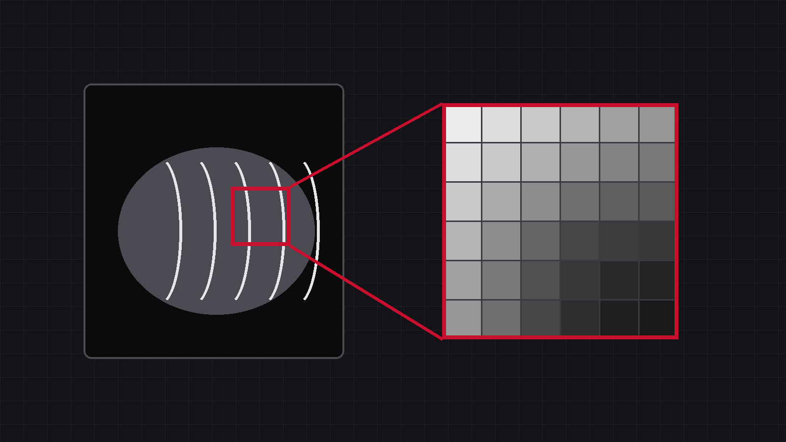
Start with a question: that X-ray image on your screen — is it a “photograph”? It isn’t. It is a table of numbers. Once that clicks, window width and level, image algorithms, even “why a JPG can never be brought back” all follow naturally.
01One radiograph = one enormous table of numbers
The sensitive surface of a flat-panel detector is divided into tiny cells — pixels. A 43 cm panel with a 139 μm pixel pitch has roughly three thousand cells along each side. At the instant of exposure, every cell records “how much X-ray reached me” as a single number.
Ten million cells, ten million numbers, laid out neatly in one table — that is the “original text” of your radiograph.
The grey you see is those numbers, translated.
Soft tissue and air
A high number means plenty of X-rays got through — little stood in their way. The lung field is the classic high-value region; on screen it translates to dark.
Bone and metal
A low number means the beam was blocked. Bone and metal implants sit in the low-value range and translate to light. Behind every grey level on your screen there is one specific number.
02The range of these numbers dwarfs your eyes
Here is the detail that matters: the range of these numbers is far wider than what your eyes can resolve. Common flat panels are 14-bit or 16-bit — each cell’s number can take up to 16,384 or 65,536 distinct levels.
The grey levels of each pixel on a 14-bit panel, against the 256 levels an ordinary monitor can display at one time — while the human eye distinguishes only a few dozen to a hundred-plus levels within a single image.
Are the tens of thousands of extra levels wasted? Quite the opposite — they are the most valuable part of the radiograph.
Picture it: among those ten million numbers, the lung field’s values are large and cluster in the high range; bone values are small and cluster in the low range. With only 256 display levels, you can “read one span at a time”. Aim those 256 levels at the high range and lung texture appears while bone washes out to solid white; aim at the low range and trabeculae emerge while the lungs sink into black.
The original never changes — only the translation does
The data are all still in that table, not one number lost — you simply choose which span to read. That is why, on the very same image, a veterinarian can drag the mouse and flip between “lung view” and “bone view”. And it is why a radiograph exported to JPG and shared over a messaging app can never be dragged back — JPG crushes 16,384 levels into 256, throwing away 98% of the numbers in the table. The original is gone.
03One step further: where image algorithms begin
If the eye can only take in 256 levels at a time, yet the table holds over ten thousand — is there a way to translate the best span for lung and the best span for bone onto one image at the same time?
There is. And it happens to be the hardest part of DR image processing — the part where the gap between systems really shows. This series will take it apart, one article at a time.
Both habits rest on the same fact: the original lives in the table, and those ten-thousand-plus levels are where the value is.
A radiograph is not a photo — it is a table.
What you see is the translation, not the original.
Source:43 cm panel / 139 μm pixels ≈ 3,093 pixels per side, based on the iLume 4343 specification; 14/16-bit = 2¹⁴ = 16,384 / 2¹⁶ = 65,536 levels; 8-bit displays = 256 levels. “The eye resolves a few dozen to a hundred-plus grey levels at once” is a common statement in visual psychophysics (it varies with luminance and viewing conditions), hence the deliberately loose figure.
IWA Medical · We build DR systems and write our own core software
Questions about veterinary DR imaging? Technical questions go straight to our engineers.