


Wired 17×17 inch flat-panel detector — 9.4 MP CsI a-Si sensor, 140 µm pixel pitch, 3.6 LP/mm spatial resolution, 4 s cycle time.
Product Overview
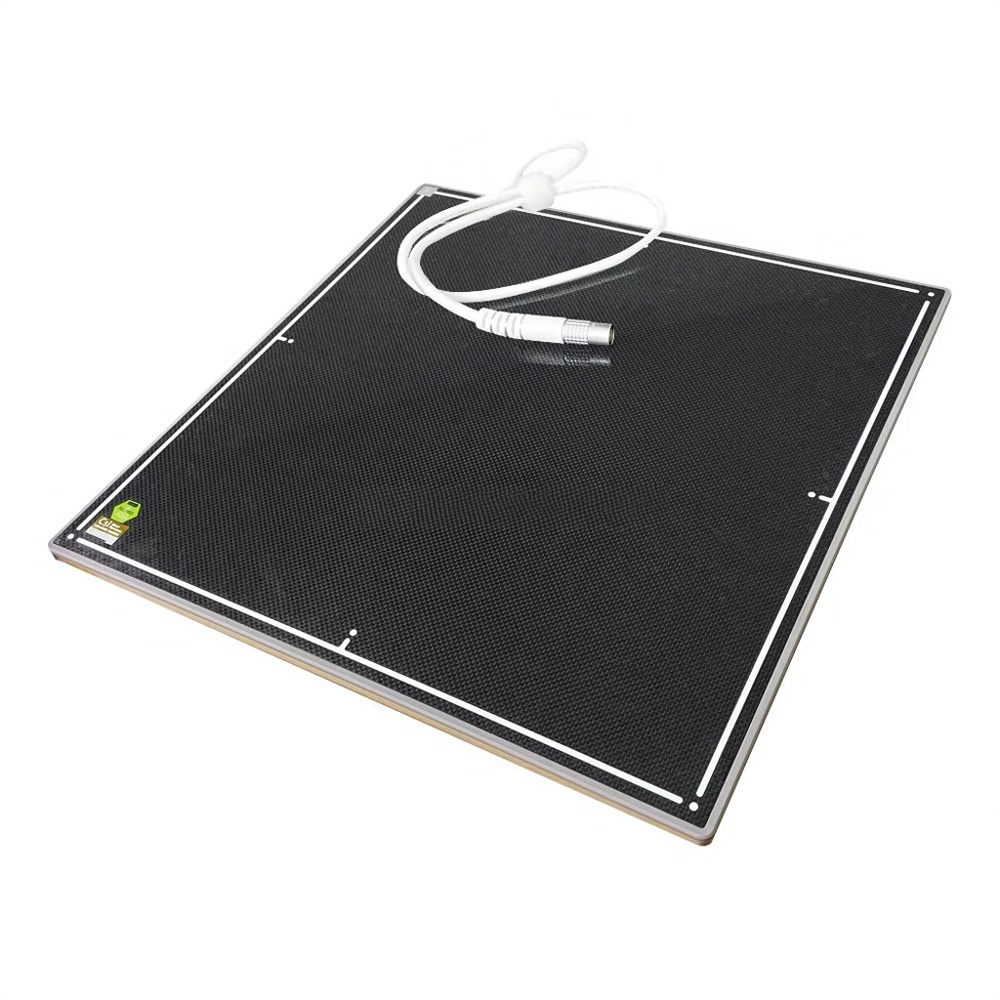
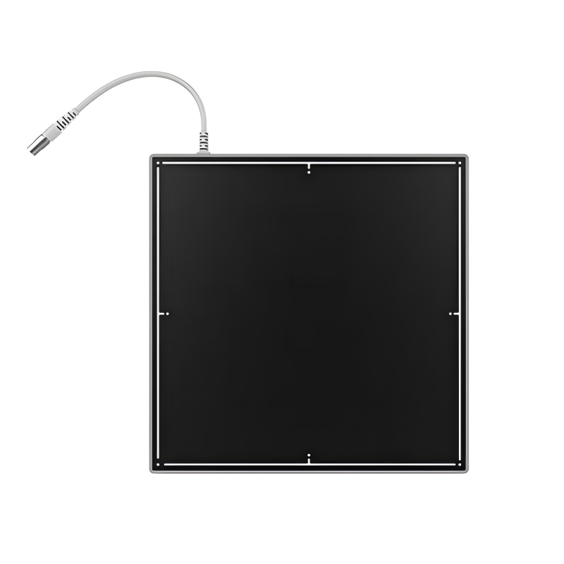
IWA-FP11 is a wired 17×17 inch flat-panel detector — 9.4 mp csi a-si sensor, 140 µm pixel pitch, 3.6 lp/mm spatial resolution, 4 s cycle time. The detector is suited for veterinary general radiography (thoracic, abdominal, skeletal), fixed-position dr upgrade for tabletop and wall-stand systems.
Key Features
- 17×17 inch active imaging area with 3072 × 3072 pixel matrix at 140 µm pitch.
- 3.6 LP/mm spatial resolution with a-Si TFT photodiode + CsI scintillator.
- 16-bit A/D conversion for full dynamic range.
- Trigger modes: AED / Prep / Software — compatible with any X-ray generator.
- Wired Ethernet data interface for stable, latency-free image transfer.
- 2 s preview image, 4 s cycle time.
- Compact 460 × 460 × 15 mm housing, 3.9 kg — fits standard ISO 4090 cassette trays for table and wall-stand mounting.
- Operating temperature 5 – 35 °C, humidity 30 – 80 % RH.
Product Gallery
Technical Specifications
| Detector technology | Amorphous silicon (a-Si) TFT |
| Scintillator | CsI |
| Active imaging area | 17 × 17 inch (432 × 432 mm) |
| Pixel matrix | 3072 × 3072 |
| Pixel pitch | 140 µm |
| Spatial resolution | 3.6 LP/mm |
| A/D conversion | 16 bit |
| Trigger modes | AED / Prep / Software |
| Preview image time | 2 s |
| Cycle time | 4 s |
| Data interface | Ethernet (wired) |
| Dimensions | 460 × 460 × 15 mm |
| Weight | 3.9 kg |
| Operating temperature | 5 – 35 °C |
| Operating humidity | 30 – 80 % RH (non-condensing) |
| Storage temperature | -10 – 55 °C |
| Storage humidity | 10 – 90 % RH (non-condensing) |
Clinical Applications
- Veterinary general radiography (thoracic, abdominal, skeletal)
- Fixed-position DR upgrade for tabletop and wall-stand systems
- Replacing legacy CR cassettes in mixed-practice clinics
- Standardised exposure workflow in equine and large-animal departments
Workflow & Installation
The detector connects via wired Ethernet to a host PC for stable, latency-free image transfer. Calibration is recommended every 6 months or when X-ray generator / tube / collimator components are replaced. Cleaning: wipe with a non-abrasive surface disinfectant; do not immerse.
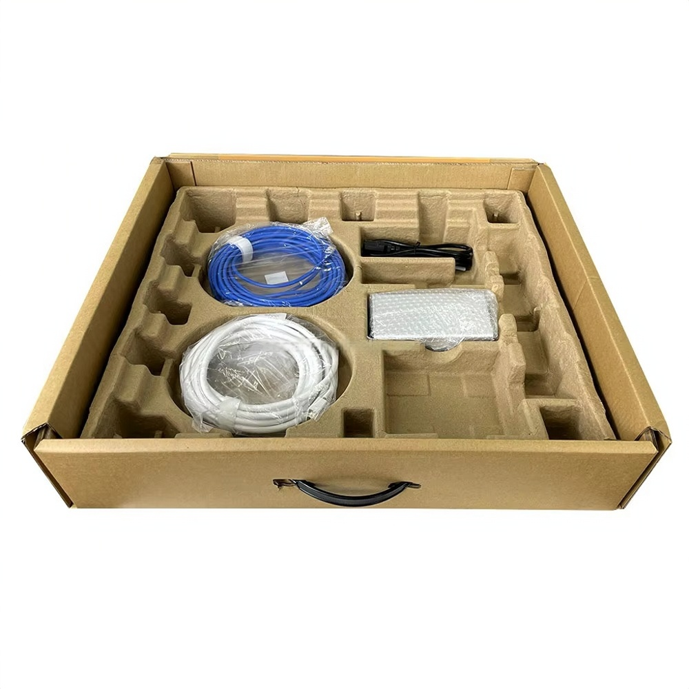
Package Contents
- 1 × IWA-FP11 flat-panel detector
- 1 × Tether cable
- 1 × Power adapter
- 1 × Acquisition software
- 1 × User manual
- 1 × Quality certificate
Why Choose IWA-FP11
- Workflow efficiency — wired Ethernet eliminates wireless reconnection issues during high-throughput clinic days.
- Clinical accuracy — 3.6 LP/mm spatial resolution and 16-bit A/D with CsI scintillator deliver diagnostic-grade image quality.
- Practice economics — standard ISO 4090 form factor enables drop-in retrofit of existing tabletop and wall-stand X-ray systems.
Warranty & Support
Manufacturer warranty per supply agreement. Spare parts and authorised in-region service available through IWA Medical / LumaVet.
FAQ
Q: Is this a static or dynamic detector?
Static. The detector is designed for single-frame radiographic exposures (not fluoroscopy or dynamic imaging).
Q: What X-ray generators is it compatible with?
Any conventional X-ray generator. The detector supports AED (Auto Exposure Detection) for use without hard-wired sync, plus software-trigger workflow.
Q: What is the data interface?
wired Ethernet, providing stable low-latency image transfer.
Q: Does it support DICOM?
The acquisition software outputs raw and processed images; DICOM 3.0 integration is typically provided by the host DR workstation software (e.g. iVision / iVet / iPet workstations) or by your existing PACS gateway.


Reviews
There are no reviews yet.